The big idea: Free ribosomes make proteins that stay in the cell; ribosomes on the rough ER make proteins that are transported in the cell or secreted.
The points to remember
- Free ribosomes float in the cytoplasm and make proteins used there, such as enzymes of glycolysis.
- The rough endoplasmic reticulum is a network of flattened membrane sacs with ribosomes attached.
- These ribosomes feed the growing polypeptide into the rough ER.
- From there the protein is carried in vesicles to the Golgi apparatus, then secreted or sent to lysosomes and membranes.
- Free and attached ribosomes are the same 80S kind; mitochondria have smaller 70S ribosomes.
Remember it as: Free: keep it. Rough ER: ship it.
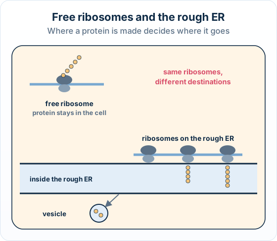
Real example: two kinds of protein: Beta cells of the pancreas make insulin on the rough ER, because insulin is secreted into the blood. Developing red blood cells make haemoglobin on free ribosomes, because it stays inside the cell.
Free preview
This is the free notes preview
You're reading the free notes. Aimnova Pro unlocks the full study experience — and you can try it with your first topic free to keep:
- FlashcardsLock in vocabulary and key terms with spaced repetition.
- Practice questionsAnswer exam-style questions and get instant AI marking.
- Mock exams & past-paper vaultSit full mocks and see exactly how examiners award marks.
- Personalised study planA daily plan built around your exam date and weak areas.
Start Studying Free Full access to Aimnova Pro · cancel anytime
The big idea: Feeding cells radioactively labelled amino acids lets you follow new proteins from organelle to organelle.
The points to remember
- The label makes the new proteins detectable, so you can track their movement.
- The organelle where the label appears first is where the protein is made: the rough ER (X).
- The label then peaks in the Golgi apparatus, and last in the secretory vesicles.
- The order of the peaks is the evidence for the route: rough ER, then Golgi, then vesicles.
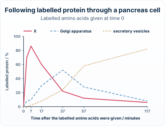
Real example: the pancreas experiment: In the 1960s James Jamieson and George Palade fed labelled amino acids to guinea pig pancreas cells and followed the label with electron microscopy, showing the route a secreted protein takes.
Know your predicted grade
Take timed mock exams and get detailed feedback on every answer. See exactly where you're losing marks.
Try Mock Exams FreeYour first topic is free to keep • No credit card required
How it is asked: Expect a labelled-amino-acid graph (identify X, explain the role of its ribosomes), a Paper 1A item on where a secreted protein is made, or an Outline of how organelles work together to secrete a protein.
IB-style questionExplain[2 marks]
Explain the role of the ribosomes located on the rough endoplasmic reticulum.
Model answer plan
See the answer broken into clear points, with guidance on how each point earns marks.
A four-mark Outline, in the Paper 2 style:
IB-style questionOutline[4 marks]
Outline how organelles interact to produce and secrete proteins.
Model answer plan
See the answer broken into clear points, with guidance on how each point earns marks.