Lipid bilayers at Higher Level: The same statement as SL. HL papers set it inside a practical: a beetroot investigation of how heat damages membranes, with questions on the method as well as the graph.
Practise this as you read
- Explain why amphipathic lipids form a continuous bilayer in water.
- Explain a graph of pigment leaking from heated beetroot.
- Outline the reason for each step of the method.
Free preview
This is the free notes preview
You're reading the free notes. Aimnova Pro unlocks the full study experience — and you can try it with your first topic free to keep:
- FlashcardsLock in vocabulary and key terms with spaced repetition.
- Practice questionsAnswer exam-style questions and get instant AI marking.
- Mock exams & past-paper vaultSit full mocks and see exactly how examiners award marks.
- Personalised study planA daily plan built around your exam date and weak areas.
To understand why a membrane is a bilayer, you have to look at a single phospholipid.
One key fact: Each phospholipid has two ends with opposite feelings about water — and that single fact is enough to make the whole bilayer form on its own.
One phospholipid is amphipathic: a hydrophilic (water-loving) phosphate head joined to two hydrophobic (water-hating) fatty-acid tails.
Interactive diagram
Explore the labelled diagram, charts and maps for this topic in full study mode.
- Hydrophilic head
- The phosphate end of a phospholipid. 'Hydrophilic' means water-loving — it is attracted to water.
- Hydrophobic tails
- The two fatty-acid ends of a phospholipid. 'Hydrophobic' means water-hating — they are repelled by water.
- Hydrophilic
- Water-loving — a part that is attracted to and mixes with water.
- Hydrophobic
- Water-hating — a part that is repelled by water and will not mix with it.
The cause → effect chain
Water on both sides
There is water on both sides of a membrane — outside the cell and inside it (the cytoplasm).
Heads face out
The hydrophilic heads are attracted to that water, so they turn to face outward on both surfaces.
Tails pushed in
The hydrophobic tails are repelled by water, so they are pushed inward, away from it, where they meet in the middle.
A bilayer forms
The result is two rows of phospholipids — a bilayer — forming spontaneously.
A water-hating core sandwiched between two water-loving surfaces
In water, phospholipids line up into a bilayer on their own: heads point outward to the water on both surfaces, tails point inward away from it.
Interactive diagram
Explore the labelled diagram, charts and maps for this topic in full study mode.
Hydrophilic heads
- Are attracted to water
- Face outward, toward the water on both surfaces
- Sit on the outer and inner edges of the membrane
- Make the membrane surfaces compatible with watery surroundings
Hydrophobic tails
- Are repelled by water
- Point inward, away from the water
- Meet in the middle to form a non-polar core
- This core blocks most polar and charged molecules from crossing
Why this core matters: Because the centre of the bilayer is hydrophobic, it acts as a barrier:
small non-polar molecules (such as oxygen and carbon dioxide) slip straight through, but large or charged/polar molecules (such as glucose and ions) cannot cross the tails freely.
That is why the membrane is selectively permeable — a direct consequence of the amphipathic phospholipids.
The mosaic of proteins: The bilayer is not bare. Scattered through it are different proteins. All these different 'tiles' make it a mosaic.
The 'tiles' of the mosaic
- Integral proteins are embedded right through the bilayer (these include the channel and carrier proteins that move substances across)
- Peripheral proteins rest on one surface
- Glycoproteins carry a carbohydrate chain on the outer surface and act as cell-recognition tags
- Cholesterol sits between the phospholipids and steadies the membrane's fluidity
| Membrane component | Where it sits | What it does |
|---|---|---|
| Phospholipid bilayer | Two rows of phospholipids forming the whole sheet | The basic structure of the membrane; controls what can cross |
| Hydrophilic heads | Facing the water on both surfaces (outside and inside) | Attracted to water, so they sit on the watery surfaces |
| Hydrophobic tails | Tucked into the middle, away from water | Repelled by water, forming a non-polar core that blocks polar molecules |
| Integral proteins (channel / carrier) | Embedded right through the bilayer | Transport molecules across; channels form pores, carriers change shape |
| Peripheral proteins | Resting on one surface of the membrane | Support, signalling and attachment roles on the membrane surface |
| Glycoproteins | Protein with a carbohydrate chain on the outer surface | Cell recognition and cell-to-cell signalling (cell 'identity tags') |
| Cholesterol | Wedged between the phospholipids | Stabilises fluidity and reduces leakiness to small molecules |
An easy way to remember: Heads love water, tails hate it. Heads point out to the water; tails hide in the middle.
Philic = friend of water (hydrophilic); phobic = afraid of water (hydrophobic).
Study smarter, not longer
Most students waste 40% of study time on topics they already know. Our AI tracks your progress and optimizes every minute.
The big idea: Put phospholipids in water and they build a bilayer by themselves. The bilayer then closes up into one continuous sheet, because that hides every tail from the water.
The points to remember
- A phospholipid is amphipathic: a hydrophilic phosphate head and two hydrophobic fatty acid tails.
- The heads form hydrogen bonds with water, so they face the water on both sides.
- The tails are attracted to each other and kept away from water, so they meet in the core.
- This happens naturally, with no energy from the cell: the bilayer assembles itself.
- An open edge would leave tails touching water, so the bilayer closes into a continuous sheet with no edges.
- Other amphipathic lipids, such as glycolipids and cholesterol, sit in the same bilayer.
Say why, not only where: Heads face the water because they are hydrophilic; tails point inwards because they are hydrophobic. A diagram with no reasons only shows where they are.
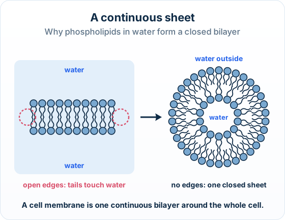
Real example: red blood cell ghosts: When red blood cells burst in pure water, the torn pieces of membrane reseal on their own into closed bubbles, called ghosts.
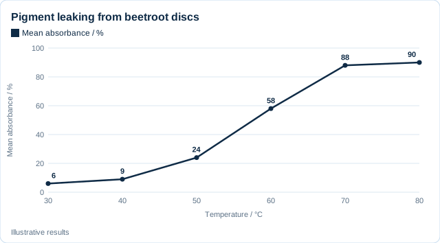
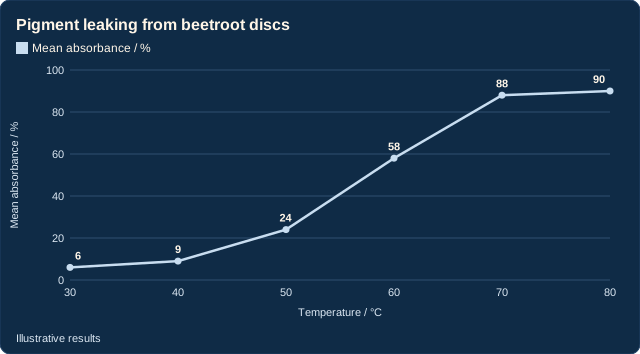
The big idea: Beetroot cells keep a red pigment inside their membranes. When heat damages the membranes, the pigment leaks out, so the redness of the water shows how much damage was done.
The points to remember
- A colorimeter measures the redness as absorbance: more pigment, higher absorbance.
- As the temperature rises, the phospholipids move more and spread apart, so gaps open in the bilayer.
- Above a certain temperature, the membrane proteins denature, leaving more gaps.
- So the membrane becomes more permeable and more pigment leaks out.
- At the highest temperatures the line levels off: all the damage has already been done.
- Anything that breaks up the bilayer or denatures its proteins, such as ethanol or extreme pH, makes the pigment leak in the same way.
Two traps: Beetroot is a plant, so do not mention cholesterol.
Do not just say the membrane becomes more fluid: say the phospholipids move apart and the proteins denature.
Practice with exam-style questions
Answer exam-style questions and get AI feedback that shows you exactly what examiners want to see in a full-marks response.
The big idea: Every step of a fair investigation has a reason. The questions on this practical ask for those reasons, one mark each.
The beetroot method and why
Equal-sized discs
Every disc has the same surface area, because surface area changes how fast pigment leaks out (diffuses).
Wash the discs
Removes the pigment from the cells that were cut open, so only leakage is measured.
Check the water bath
Read a thermometer or temperature probe at intervals throughout the 30 minutes.
Repeat at each temperature
Gives a reliable mean and lets anomalies (odd results) be spotted.
Use a colorimeter
It is objective (no human judgement) and gives precise values between the points of a colour scale.
Words that score nothing: Repeats do not make results more accurate, and a colorimeter is not just more accurate: say reliable for repeats and precise or objective for the colorimeter.
How this is tested: Membrane structure comes up as a Paper 2 Explain or Outline, a Paper 1 diagram question, and model comparisons.
| Where | What you are asked | What scores |
|---|---|---|
| Paper 2 (3–4 marks, Explain or Outline), a regular | Explain how the amphipathic properties of phospholipids allow a bilayer to form | Heads to the water, tails away from it, bilayer forms spontaneously |
| Paper 1 (multiple choice) | A labelled fluid-mosaic diagram: identify which regions are hydrophilic, which component does cell recognition, or name labelled components X and Y | Cell recognition = the glycoprotein |
Exam-style question — explain how amphipathic phospholipids form a bilayer
Explain how the amphipathic nature of phospholipids allows them to form the bilayer of a cell membrane. [4]
How to score all four marks
- Define amphipathic. Each phospholipid is amphipathic — it has a hydrophilic (water-loving) head and hydrophobic (water-hating) tails.
- Place the heads. There is water on both sides of the membrane, so the hydrophilic heads face outward toward the water on each surface.
- Place the tails. The hydrophobic tails are repelled by water, so they are pushed inward, away from the water, meeting in the middle.
- State the outcome. This produces two rows of phospholipids — a bilayer — which forms spontaneously in water, with a hydrophobic core between two hydrophilic surfaces. (Award 1 mark each for: amphipathic / heads + tails; heads to the water; tails away from water; bilayer forms.)
Final answer
Phospholipids are amphipathic; the hydrophilic heads face the water on both surfaces and the hydrophobic tails point inward away from water, so two rows line up spontaneously into a bilayer with a hydrophobic core.
Why this scores full marks: Each step is a separate scoring point and the answer is a clear cause → effect chain: amphipathic structure causes the heads-out, tails-in arrangement, which causes the bilayer.
A common slip is to describe the bilayer without explaining that water on both sides is what pulls the heads out and pushes the tails in.
A three-mark Explain on a graph, in the Paper 1B style:
Discs of red cabbage leaf were kept in water at different temperatures. The red colour of the water stayed low up to 45 °C, rose steeply between 50 and 65 °C, then levelled off. Using your knowledge of membrane structure, explain these results.
Model answer plan
See the answer broken into clear points, with guidance on how each point earns marks.