aımnova
DashboardMy LearningCrash CoursesStudy Plan

Aimnova site navigation

Stay in the loop

Get the latest study resources and updates

New features, study tips and exam insights — straight to your inbox.

IB Diploma

  • IB Past Papers
  • IB Study Notes
  • IB Question Bank
  • IB Mock Exams
  • IB Revision

IB Subjects

  • IB Math AA
  • IB Math AI
  • IB Economics
  • IB Business Management
  • IB Physics
  • IB Biology
  • View all IB subjects→

IB Past Papers

  • IB Math AA HL Past Papers
  • IB Math AA SL Past Papers
  • IB Math AI HL Past Papers
  • IB Math AI SL Past Papers
  • IB Economics HL Past Papers
  • IB Economics SL Past Papers
  • IB ESS Past Papers
  • View all past papers→

Study Resources

  • Study Notes
  • Question Bank
  • Mock Exams
  • Crash Courses
  • Flashcards
  • Revision Guide
  • Exam Skills
  • Command Terms
  • Grade Calculator
  • Exam Timetable 2026

Aimnova

  • Features
  • Pricing
  • For Teachers
  • For Schools
  • For Parents
  • About Us
  • Blog
  • Contact
aımnova

IB study, practice and feedback built around the real exam.

TermsPrivacyCookies·© 2026 Aimnova. All rights reserved.10c8990

Aimnova is not affiliated with or endorsed by the International Baccalaureate Organization (IB).

NotesBiology HLTopic 1.2Structure of a nucleosome
Back to Biology HL Topics
1.2.134 min read

Structure of a nucleosome (Biology HL)

IB Biology · Unit 1

Exam preparation

Practice the questions examiners actually ask

Our question bank mirrors real IB exam papers. Practice under timed conditions and track your progress across topics.

Start Practicing

Contents

  1. 1The structure of a nucleosome
  2. 2Reading a computer model of a nucleosome
  3. 3Exam-style question (step by step)
The big idea: In eukaryotes, DNA is wound around balls of protein called histones. One ball with its DNA is a nucleosome: the basic unit of packing in a chromosome.

The points to remember

  • A nucleosome is a length of DNA wrapped around a core of eight histone proteins.
  • The core is a ball of eight histones, called an octamer: four types of histone, two of each.
  • The DNA double helix wraps around the outside of the core almost twice.
  • A ninth histone (H1) binds where the DNA enters and leaves, holding the DNA on the core.
  • Linker DNA joins one nucleosome to the next, like the string between beads.
  • Nucleosomes help pack long DNA molecules into the nucleus. Only eukaryotes have them: bacterial DNA is naked.
Remember it as: Eight in the bead, one on the string.
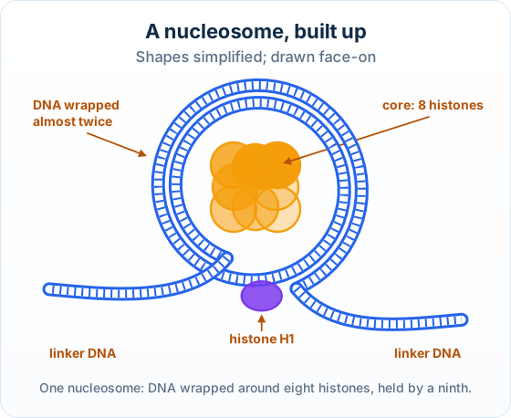
A nucleosome drawn face-on: a ball of eight histones in four shades, DNA wrapped around it almost twice, a histone H1 where the DNA enters and leaves, and linker DNA running off to each side
Eight histones, DNA wrapped almost twice, a ninth histone, linker DNA.
Real example: the nucleosome in atomic detail: In 1997 Karolin Luger and her colleagues used X-ray crystallography to work out the nucleosome core atom by atom: 146 base pairs of DNA wrapped about 1.65 times around the eight histones.

Free preview

This is the free notes preview

You're reading the free notes. Aimnova Pro unlocks the full study experience — and you can try it with your first topic free to keep:

  • FlashcardsLock in vocabulary and key terms with spaced repetition.
  • Practice questionsAnswer exam-style questions and get instant AI marking.
  • Mock exams & past-paper vaultSit full mocks and see exactly how examiners award marks.
  • Personalised study planA daily plan built around your exam date and weak areas.
Start Studying Free Full access to Aimnova Pro · cancel anytime
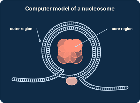
The big idea: Computer models let you turn a nucleosome round, zoom in and colour its parts. Exam questions show such a model and ask what each region is, or what the software is good and bad at.

The points to remember

  • Molecular visualization software builds a 3-D model from data on the position of every atom.
  • The DNA shows as a double helix on the outside, wrapped round the core.
  • The core shows as proteins: you can see their alpha helices, and colouring by chain shows the eight histones.
  • Histones are positively charged and the DNA's phosphates are negatively charged, so the two attract and hold together.
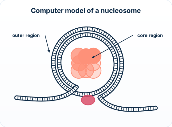
A computer-style model of a nucleosome with the outer region (the wrapped DNA) and the core region (the histones) labelledA computer-style model of a nucleosome with the outer region (the wrapped DNA) and the core region (the histones) labelled
Outer region: DNA. Core region: eight histone proteins.

Benefits of the software

  • Shows a structure too small to see directly
  • Can be rotated to see it in 3-D from any angle
  • Can zoom in on one part, or colour parts to tell them apart

Drawbacks

  • It is a model: simplified, so some detail is left out
  • It rests on assumptions and may not match the real structure exactly
  • It can be costly to make or run
Try it yourself: On the RCSB Protein Data Bank website, search for the structure 1KX5 (a nucleosome core) and open the 3-D view. Rotate it to find the DNA outside and the histones inside.

Get feedback like a real examiner

Submit your answers and get instant feedback — what you did well, what's missing, and exactly what to write to score full marks.

Try AI Tutor FreeYour first topic is free to keep • No credit card required
How it is asked: Paper 1A: what a nucleosome is, or labels on a diagram. Paper 1B, on a computer model: identify a region with a reason, describe the core or the whole nucleosome, or give a benefit and a drawback of the software.
IB-style questionDescribe[3 marks]

The image of a nucleosome was made using molecular visualization software. Describe the structure of the nucleosome.

Model answer plan

See the answer broken into clear points, with guidance on how each point earns marks.

Claim your free topic

The skill question that goes with the model:

IB-style questionSuggest[2 marks]

Suggest one benefit and one disadvantage of using molecular visualization to study a nucleosome.

Model answer plan

See the answer broken into clear points, with guidance on how each point earns marks.

Claim your free topic

Try an IB Exam Question — Free AI Feedback

Test yourself on Structure of a nucleosome. Write your answer and get instant AI feedback — just like a real IB examiner.

The DNA of the bacterium Escherichia coli is not organized into nucleosomes.

a reason for this.
[1 mark]

Related Biology HL Topics

Continue learning with these related topics from the same unit:

1.1.1Water as the medium for life1.1.2Hydrogen bonds from the polar covalent bonds in water1.1.3Cohesion of water and its consequences for organisms1.1.4Adhesion of water to polar or charged materials
View all Biology HL topics
Improve your exam techniqueCommand terms, paper structure, and mark-scheme tips for Biology HL
Previous1.2.12Purine-to-pyrimidine bonding and the stability of the DNA helixNextEvidence from the Hershey–Chase experiment for DNA as the genetic material1.2.14

21 practice questions on Structure of a nucleosome

Students who practiced this topic on Aimnova scored 82% on average. Try free practice questions and get instant AI feedback.

Try 3 Free QuestionsView All Biology HL Topics