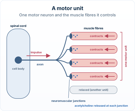
The big idea: Skeletal muscle is controlled in motor units: one motor neuron and all the muscle fibres it supplies. They always contract together.
The points to remember
- A motor neuron has its cell body in the spinal cord; its long axon runs to the muscle.
- The axon branches; each branch ends on one muscle fibre at a neuromuscular junction.
- At each junction acetylcholine is released; it triggers an impulse in the muscle fibre.
- So every fibre in the unit contracts at once. Each fibre belongs to one unit only.
- A stronger contraction recruits more motor units; small units give fine control.
Small motor units
- Few fibres per motor neuron
- Small, precise movements
- Eye and finger muscles
Large motor units
- Many fibres per motor neuron
- Powerful movements
- Thigh and calf muscles
Remember it as: One neuron, its fibres, one junction each: they fire together.
Real example: eye v calf. A motor unit in a muscle that moves the eye controls only about ten fibres, so the eye moves very precisely. One in the calf muscle controls up to about two thousand fibres, for power.
Trap: A motor unit is not one muscle fibre and not the whole muscle: it is one motor neuron plus all the fibres it controls.
Free preview
This is the free notes preview
You're reading the free notes. Aimnova Pro unlocks the full study experience — and you can try it with your first topic free to keep:
- FlashcardsLock in vocabulary and key terms with spaced repetition.
- Practice questionsAnswer exam-style questions and get instant AI marking.
- Mock exams & past-paper vaultSit full mocks and see exactly how examiners award marks.
- Personalised study planA daily plan built around your exam date and weak areas.
The big idea: In the sliding-filament model, myosin heads pull on actin to shorten the muscle. But a muscle must only contract when it is told to, so there has to be a switch.
The switch is calcium: That switch is calcium (Ca²⁺). Two thin-filament proteins do the gatekeeping.
Tropomyosin
- A thread that lies along the actin.
- It physically covers the myosin-binding sites.
Troponin
- A small protein clamped onto tropomyosin that holds it in place.
- It carries the Ca²⁺ binding site.
From rest to contraction
At rest
The sites are blocked, so myosin cannot attach.
Nerve impulse
A nerve impulse floods the cell with Ca²⁺.
Block lifted
The block is lifted, and contraction begins.
The cross-bridge cycle is the engine behind sliding-filament contraction: myosin heads grab the exposed sites on actin and pull the thin filaments toward the centre, shortening the sarcomere.
Interactive diagram
Explore the labelled diagram, charts and maps for this topic in full study mode.
- Actin (thin filament)
- The thin filament that myosin pulls on; it carries the myosin-binding sites.
- Myosin (thick filament)
- The thick filament whose heads attach to actin and perform the power stroke.
- Tropomyosin
- A thread-like protein that lies along actin and BLOCKS the myosin-binding sites when the muscle is at rest.
- Troponin
- A protein attached to tropomyosin; it holds tropomyosin in the blocking position and has a binding site for Ca²⁺.
- Calcium (Ca²⁺)
- The ion released on stimulation; it binds troponin and acts as the switch that exposes the binding sites.
- Sarcoplasmic reticulum
- The internal store of Ca²⁺ in a muscle fibre; it releases Ca²⁺ on a nerve impulse and pumps it back to end contraction.
- Cross-bridge
- A temporary link formed when a myosin head attaches to an exposed binding site on actin.
Don't confuse the two 'trop-' proteins: Tropomyosin = the long thread that covers the binding sites (it does the blocking).
Troponin = the small clamp that holds tropomyosin and binds Ca²⁺ (it does the sensing).
An easy way to remember: troponin binds the ion (Ca²⁺); tropomyosin sits over the myosin sites.
Stop wasting time on topics you know
Our AI identifies your weak areas and focuses your study time where it matters. No more overstudying easy topics.
How this is tested: Questions ask how a motor neuron connects to muscle fibres, and what the calcium switch and the cross-bridge cycle then do.
Common Outline / Explain questions
- The role of calcium ions in starting contraction: Ca²⁺ → troponin → tropomyosin moves → sites exposed.
- The role of ATP: binds to detach the head; hydrolysed to re-cock it.
- A longer Explain asks you to put the whole molecular cycle in order.
Score points for each step, in order
Blocked sites at rest
Tropomyosin covers the binding sites.
Ca²⁺ released
Then Ca²⁺ binds troponin.
Tropomyosin moves
The binding sites are exposed.
Myosin binds
A cross-bridge forms.
Power stroke
The head pulls actin.
ATP binds
The head detaches.
ATP hydrolysed
The head is re-cocked, and the cycle repeats.
Watch the role-split: Don't say ATP opens the binding sites (that's Ca²⁺), and don't say Ca²⁺ powers the power stroke (the energy comes from ATP).
Exam-style question — the molecular roles of Ca²⁺ and ATP in contraction
Explain the roles of calcium ions and ATP in the contraction of a muscle fibre. [6]
How to score all six marks
- Blocked at rest. At rest, tropomyosin (held by troponin) covers the myosin-binding sites on actin, so myosin cannot attach.
- Ca²⁺ released and binds troponin. On stimulation, Ca²⁺ is released from the sarcoplasmic reticulum and binds to troponin.
- Sites exposed (Ca²⁺ = the switch). Troponin changes shape and moves tropomyosin away, so the myosin-binding sites are exposed — this is calcium's role: the switch that allows cross-bridges to form.
- Cross-bridge + power stroke. Myosin heads bind the exposed sites (cross-bridges) and the power stroke pulls actin toward the centre of the sarcomere.
- ATP detaches the head. A new ATP molecule binds the myosin head, breaking the cross-bridge so the head detaches from actin.
- ATP re-cocks the head. The ATP is hydrolysed (ATP → ADP + Pi); the energy re-cocks the head for the next cycle — so ATP's role is detachment and re-cocking, and the cycle repeats while Ca²⁺ and ATP are present.
Final answer
At rest tropomyosin (held by troponin) blocks the binding sites. Ca²⁺ released from the sarcoplasmic reticulum binds troponin, moving tropomyosin so the sites are exposed (Ca²⁺ = the switch). Myosin heads bind and the power stroke pulls actin inward. A new ATP binds the head to detach it, then ATP is hydrolysed to re-cock it (ATP = energy for detachment and re-cocking). The cycle repeats while Ca²⁺ and ATP are present.
Why this scores full marks: It keeps the roles separate: Ca²⁺ as the switch (binds troponin, exposes the sites) and ATP as the energy (binds to detach, then is hydrolysed to re-cock). It also puts the molecular steps in the right order.
A common way to lose marks: A common way to lose marks is to mix the roles up: saying ATP exposes the binding sites, or that Ca²⁺ powers the power stroke.
| Calcium (Ca²⁺) | ATP | |
|---|---|---|
| What it is | An ion released from the sarcoplasmic reticulum on stimulation | The cell's energy molecule, hydrolysed to ADP + Pi |
| What it binds | Troponin | The myosin head |
| Its job | The SWITCH — controls whether the binding sites are exposed | The ENERGY — powers detachment and re-cocking of the head |
| When missing… | Tropomyosin re-blocks the sites → muscle relaxes | The myosin head cannot let go → it stays stuck (rigor) |
A 3-mark Outline, in the papers' style:
Outline the structure of a motor unit in a skeletal muscle.
Model answer plan
See the answer broken into clear points, with guidance on how each point earns marks.