The big idea: Striated (skeletal) muscle looks striped under the microscope because it is built from millions of identical repeating units lined up end to end.
The sarcomere
- One of these repeating units is a sarcomere: the functional contractile unit of muscle.
- A sarcomere is defined as the region between two adjacent Z-discs.
- When the muscle contracts, every sarcomere shortens a little, and together that shortens the whole fibre.
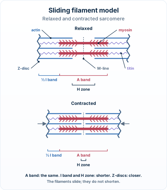
One sarcomere lies between two Z-discs. Thin ACTIN filaments are anchored to the Z-discs; thick MYOSIN filaments sit in the centre, held in register by the M-line. The light I band is actin only, the dark A band is the full length of myosin, and the H zone is the central myosin-only region.
Interactive diagram
Explore the labelled diagram, charts and maps for this topic in full study mode.
- Sarcomere
- The functional contractile unit of striated muscle — the region between two adjacent Z-discs.
- Z-disc
- The protein boundary at each end of a sarcomere; the actin filaments are anchored to it.
- Thin filament (actin)
- A thin protein filament anchored to the Z-discs and projecting inward toward the centre of the sarcomere.
- Thick filament (myosin)
- A thick protein filament that carries protruding heads (cross-bridges); it sits in the centre of the sarcomere.
- M-line
- The line down the middle of the sarcomere that links the myosin filaments and holds them in register.
- Titin
- A giant elastic protein that anchors myosin to the Z-disc, keeps it centred, and provides passive recoil (elasticity).
Thin = actin, thick = myosin: Thin filaments are actin — anchored to the Z-discs at the ends.
Thick filaments are myosin — in the centre, with heads that stick out.
An easy way to remember: myosin = the middle, massive (thick) filament with the moving heads.
Free preview
This is the free notes preview
You're reading the free notes. Aimnova Pro unlocks the full study experience — and you can try it with your first topic free to keep:
- FlashcardsLock in vocabulary and key terms with spaced repetition.
- Practice questionsAnswer exam-style questions and get instant AI marking.
- Mock exams & past-paper vaultSit full mocks and see exactly how examiners award marks.
- Personalised study planA daily plan built around your exam date and weak areas.
A sarcomere is built from just a few proteins, each in a fixed place.
Once you know where each one sits, the striped banding you see under the electron microscope (EM) follows automatically. The bands are simply named after which filaments lie there and whether they overlap.
What's inside one sarcomere
- Z-discs at each end form the boundary; actin (thin) filaments are anchored to them and reach inward.
- Myosin (thick) filaments sit in the centre, carrying protruding heads (cross-bridges) that point toward the actin.
- The M-line runs down the middle and links the myosin filaments, keeping them lined up.
- Titin, a giant elastic protein, runs from each Z-disc to the myosin, anchoring it and acting like a molecular spring.
| Component | What it is | Where it sits | Its job |
|---|---|---|---|
| Actin (thin filament) | A thin protein filament | Anchored to the Z-discs, projecting inward toward the centre | The filament the myosin heads pull on during contraction |
| Myosin (thick filament) | A thick protein filament with protruding HEADS (cross-bridges) | In the centre of the sarcomere, held in register by the M-line | Its heads grab and pull the actin to shorten the sarcomere |
| Titin | A giant ELASTIC protein (a molecular spring) | Runs from the Z-disc to the myosin filament | Anchors myosin to the Z-disc, keeps it centred, and provides recoil / elasticity |
| Z-disc | A protein plate / boundary line | At each END of the sarcomere | Anchors the actin filaments; marks where one sarcomere meets the next |
| M-line | A protein line down the middle | The centre of the sarcomere | Links the myosin filaments and holds them in register |
Titin — the molecular spring: Titin is the largest protein in the body, and it does three jobs at once.
Titin's three jobs
- Anchors each myosin filament to the Z-disc, so myosin can't drift out of place.
- Centres the myosin in the middle of the sarcomere (keeps it in register with the M-line).
- Provides recoil: like a stretched spring, titin pulls the sarcomere back to its resting length after a stretch and resists overstretching. This passive elasticity is why a relaxed muscle springs back rather than staying floppy.
Now the banding. The bands are just labels for regions of the sarcomere, based on what lies there:
Reading the bands (light vs dark)
- I band — actin only (no myosin), so it looks light. It spans a Z-disc.
- A band — the full length of the myosin filament. Because myosin is present (and overlaps actin at its edges), it looks dark. The A band is as wide as one whole myosin filament.
- H zone — the myosin-only region in the middle of the A band, where no actin reaches. It's a paler stripe inside the dark A band.
- M line — the centre line that links the myosin filaments (sits in the middle of the H zone).
- Z disc — the boundary of the sarcomere, in the middle of the I band, where actin is anchored.
Reading the bands: I band = actin only (light, across each Z-disc) · A band = the full myosin length (dark) · H zone = myosin only in the middle (no actin overlap) · M line = centre · Z-disc = sarcomere boundary.
Interactive diagram
Explore the labelled diagram, charts and maps for this topic in full study mode.
| Region | What lies there | Appearance under EM | Quick definition |
|---|---|---|---|
| I band | Actin only (no myosin) | Light | The light band; spans a Z-disc |
| A band | The full length of the myosin filament (incl. the actin–myosin overlap) | Dark | As wide as one whole myosin filament |
| H zone | Myosin only (the middle of the A band, no actin overlap) | Paler centre of the dark A band | Myosin with no actin |
| M line | The centre, linking the myosin filaments | A line in the middle of the H zone | Centre / register of the myosin |
| Z disc | Where actin is anchored | A dark line in the middle of the I band | The sarcomere boundary |
The logic in one line: Light = actin only (I band), dark = myosin present (A band), and the myosin-only middle = H zone.
Every band name is just a statement about which filament is there — that's all you need to remember to label a sarcomere diagram correctly.
Know your predicted grade
Take timed mock exams and get detailed feedback on every answer. See exactly where you're losing marks.
The big idea: A muscle shortens, but the protein filaments inside it do not. This is the sliding filament model.
Thin filaments
- Made of actin.
- Anchored to plates called Z-discs.
Thick filaments
- Made of myosin.
- Sit in the centre.
What happens in contraction: Inside each muscle fibre, repeating units called sarcomeres contain both kinds of filament. During contraction the actin and myosin filaments slide past each other so that they overlap more. The filaments keep the same length; only the amount of overlap changes.
One sarcomere lies between two Z-discs. RELAXED (top) vs CONTRACTED (bottom): the actin and myosin filaments keep the same length but slide so they OVERLAP MORE — the Z-discs come closer, and the I band and H zone shrink while the A band stays the same.
Interactive diagram
Explore the labelled diagram, charts and maps for this topic in full study mode.
- Sarcomere
- The repeating contractile unit of a muscle fibre, running from one Z-disc to the next.
- Z-disc
- The plate at each end of a sarcomere; the thin actin filaments are anchored to it.
- Actin (thin filament)
- The thin protein filament anchored to the Z-discs and projecting inward toward the centre.
- Myosin (thick filament)
- The thick protein filament in the centre of the sarcomere; it pulls on the actin.
- Sliding filament model
- The model that explains contraction: actin and myosin slide past each other so they overlap more, and the sarcomere shortens — the filaments do not shorten.
Slide, not shrink: The single most-tested point: the filaments slide, they do not shorten.
A sarcomere gets shorter because the overlap increases, just like two combs pushed into each other take up less space without either comb getting shorter.
A sarcomere has a striped banding pattern you can name. Because contraction only increases overlap, each band changes in a fixed, predictable way.
You can work each one out from where actin and myosin sit. Read it as structure → observation: think about what overlaps with what, and the band change follows.
What changes when the sarcomere contracts
- Myosin pulls the actin filaments inward, toward the centre (the M-line).
- Because the actin is anchored to the Z-discs, the Z-discs are pulled closer together — so the sarcomere shortens.
- There is now more overlap between actin and myosin.
- The I band (thin actin only) gets shorter — more of the actin is now overlapped by myosin.
- The H zone (thick myosin only) gets shorter — the actin tips slide inward and cover more of the myosin.
- The A band (the full myosin length) stays the same — myosin does not change length.
Read the bands to spot contraction: compare top (relaxed) and bottom (contracted). Z-to-Z (sarcomere) ↓, I band ↓, H zone ↓ — but the A band (the full myosin length) is UNCHANGED, because myosin does not shorten.
Interactive diagram
Explore the labelled diagram, charts and maps for this topic in full study mode.
The A band is your control: The A band equals the length of the thick myosin filaments.
Since myosin never shortens, the A band never changes length — not when relaxed, not when fully contracted.
This is the proof that the filaments slide rather than shrink: if the filaments themselves got shorter, the A band would shrink too. It doesn't, so they must be sliding.
| Region | What it is | During contraction | Why |
|---|---|---|---|
| Sarcomere (Z to Z) | The unit between two Z-discs | SHORTER | Filaments slide inward, pulling the Z-discs closer |
| Z-discs | The plates the actin filaments are anchored to | Pulled CLOSER together | Myosin pulls the actin (and so the Z-discs) toward the centre |
| I band | The region with thin actin only (no myosin overlap) | SHORTER | More overlap means less actin is left uncovered by myosin |
| H zone | The central region with thick myosin only (no actin overlap) | SHORTER | Actin slides inward toward the M-line, covering more of the myosin |
| A band | The full length of the thick myosin filaments | UNCHANGED | Myosin keeps its length — the A band always equals it |
One sarcomere → the whole muscle: Each muscle fibre is a long chain of sarcomeres joined end to end (in series).
When every sarcomere shortens by a small amount at the same time, the shortenings add up along the fibre, so the whole fibre — and the whole muscle — shortens noticeably.
So a big movement comes from many tiny sliding events happening together in series.
Draw it like this: the same sarcomere relaxed and contracted, with the bands marked.
Get feedback like a real examiner
Submit your answers and get instant feedback — what you did well, what's missing, and exactly what to write to score full marks.
Read the mechanism as a chain of cause and effect. It starts with the sites blocked, and the Ca²⁺ switch flips them open.
Then the myosin head runs a repeating four-step cross-bridge cycle as long as Ca²⁺ and ATP are both present.
Turning the switch ON
- At rest: tropomyosin (held by troponin) lies over the actin and blocks the myosin-binding sites, so no cross-bridges can form.
- A nerve impulse reaches the fibre and Ca²⁺ is released from the sarcoplasmic reticulum.
- Ca²⁺ binds troponin, which changes shape and pulls tropomyosin away from the binding sites.
- The myosin-binding sites are now EXPOSED — the cross-bridge cycle can run.
| At rest (relaxed) | When stimulated (contracting) | |
|---|---|---|
| Ca²⁺ level in the cytoplasm | Low — Ca²⁺ is stored in the sarcoplasmic reticulum | High — Ca²⁺ released from the sarcoplasmic reticulum |
| Troponin | Holds tropomyosin in the blocking position | Ca²⁺ binds it → it changes shape and shifts tropomyosin |
| Tropomyosin | Covers (BLOCKS) the myosin-binding sites on actin | Moved AWAY → binding sites are EXPOSED |
| Myosin heads | Cannot attach — no cross-bridges form | Attach to actin → cross-bridges + power strokes |
| Outcome | No sliding — muscle stays relaxed | Filaments slide → sarcomere shortens (contracts) |
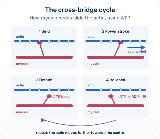
One cross-bridge cycle (repeats)
- Bind: a myosin head attaches to an exposed site on actin, forming a cross-bridge.
- Power stroke: the head pivots and pulls the actin filament toward the centre of the sarcomere — this is the stroke that shortens the muscle.
- Detach: a new ATP binds the myosin head, which breaks the cross-bridge so the head lets go of actin.
- Re-cock: the ATP is hydrolysed (ATP → ADP + Pi); the energy released re-cocks the head back to its 'ready' position, set for the next cycle.
| Step | What happens | What is used / needed |
|---|---|---|
| 1. Bind | The myosin head attaches to the exposed site on actin — a cross-bridge | Sites already exposed (needs Ca²⁺) |
| 2. Power stroke | The head pivots, pulling the actin filament toward the sarcomere centre | Energy stored from the previous ATP hydrolysis |
| 3. Detach | A new ATP molecule binds the head, breaking the cross-bridge so it lets go | 1 ATP binds |
| 4. Re-cock | ATP is hydrolysed (ATP → ADP + Pi); the head returns to its 'ready' position | ATP hydrolysed — energy released |
The two jobs: Ca²⁺ is the switch, ATP is the energy: Keep the two molecules' roles separate: questions often test this. Ca²⁺ unlocks the door; ATP does the work of letting go and resetting.
Ca²⁺ = the switch
- It binds troponin and decides whether the binding sites are open.
- It controls if the cycle can run, not the movement itself.
ATP = the energy
- It is needed twice in each cycle.
- One ATP binds to detach the head.
- The same ATP is then hydrolysed to re-cock the head for the next pull.
| Calcium (Ca²⁺) | ATP | |
|---|---|---|
| What it is | An ion released from the sarcoplasmic reticulum on stimulation | The cell's energy molecule, hydrolysed to ADP + Pi |
| What it binds | Troponin | The myosin head |
| Its job | The SWITCH — controls whether the binding sites are exposed | The ENERGY — powers detachment and re-cocking of the head |
| When missing… | Tropomyosin re-blocks the sites → muscle relaxes | The myosin head cannot let go → it stays stuck (rigor) |
How the muscle relaxes: When the nerve impulse stops, the Ca²⁺ is actively pumped back into the sarcoplasmic reticulum. With Ca²⁺ gone, troponin returns to its original shape, tropomyosin slides back over the binding sites, and no new cross-bridges form. The muscle relaxes.
Rigor mortis: This is why a dead body goes stiff (rigor mortis): with no ATP left, the heads cannot detach from actin, so the cross-bridges stay locked.
The cycle in four pictures:
Ready to ace Sliding filament model of muscle contraction?
You've seen the preview. Here's what you'll unlock:
Bite-Sized Study Notes
Full syllabus broken into chunks that stick
Exam Practice
See exactly how to score full marks
AI Feedback
Know exactly what examiners want
Smart Study Plan
Know exactly what to study next