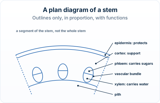
Dicot stems at Higher Level: The same statement as SL. HL papers pair stem micrographs with naming a tissue and its function, and with plan diagrams.
Practise this as you read
- Draw a labelled plan diagram of a stem section.
- Identify a labelled tissue and its function.
Free preview
This is the free notes preview
You're reading the free notes. Aimnova Pro unlocks the full study experience — and you can try it with your first topic free to keep:
- FlashcardsLock in vocabulary and key terms with spaced repetition.
- Practice questionsAnswer exam-style questions and get instant AI marking.
- Mock exams & past-paper vaultSit full mocks and see exactly how examiners award marks.
- Personalised study planA daily plan built around your exam date and weak areas.
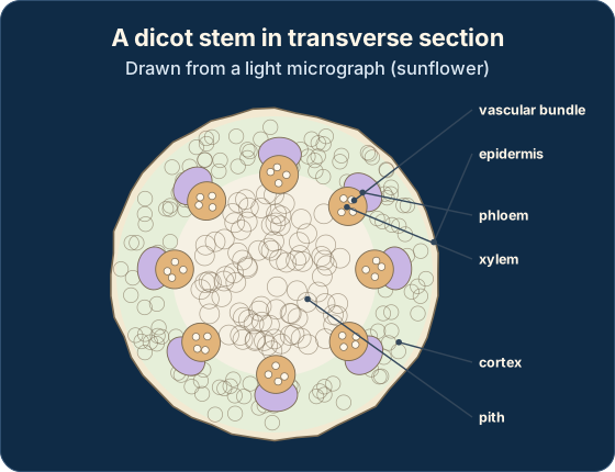
The big idea: In a dicot stem the vascular bundles form a ring, between the cortex outside and the pith in the centre.
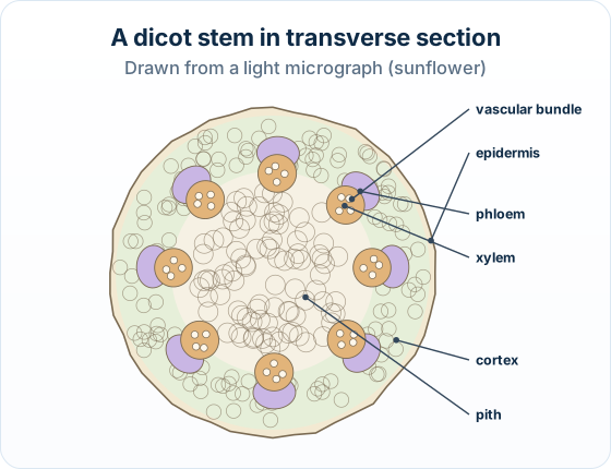
| Tissue | Where | Main function |
|---|---|---|
| Epidermis | One thin outer layer | Protects the stem; cuts water loss |
| Cortex | Wide band under the epidermis | Support and storage |
| Vascular bundle | In a ring, between cortex and pith | Transport and support |
| Phloem | Outer side of each bundle | Carries sugars and amino acids |
| Xylem | Inner side of each bundle | Carries water and minerals; support |
| Pith | The centre | Storage |
The points to remember
- Order from the outside: epidermis, cortex, vascular bundles, pith.
- In each bundle, phloem faces outwards and xylem faces inwards, towards the centre.
- Phloem carries sugars and amino acids, not 'food' or 'nutrients'.
Remember it as: Phloem out, xylem in.
Real example: the sunflower. Young sunflower stems are the classic school slide: separate vascular bundles in a ring, each with phloem towards the outside and xylem towards the centre.
Learn what examiners really want
See exactly what to write to score full marks. Our AI shows you model answers and the key phrases examiners look for.
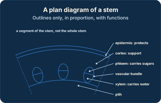
What a plan diagram is: A plan diagram shows where each tissue is, as outlines. It never shows cells.
The points to remember
- Draw the segment you are asked for (area X), not the whole stem, with more than one vascular bundle.
- Outline the epidermis, the cortex under it, each vascular bundle and the pith, in proportion.
- Split each bundle: phloem on the outer side, xylem on the inner side.
- Label with ruled lines; add a short function for each tissue when asked to annotate.
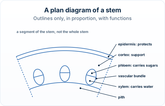
Three traps: Drawing cells, or the whole stem when only part is asked for, loses marks.
The cortex label must point to the band just under the epidermis.
A leaf section is not a stem section: leaves have palisade and spongy layers.
How it is asked: Expect a stem micrograph: draw a plan of part of it, identify the tissue that carries water, or state what the phloem does.
The micrograph shows part of a sunflower stem in transverse section. Draw a labelled plan diagram to show the distribution of tissues.
Model answer plan
See the answer broken into clear points, with guidance on how each point earns marks.