The fluid mosaic model at Higher Level: The same statement as SL. At HL the drawing is the starting point for membrane fluidity: how fatty acids and cholesterol set how fluid the membrane is.
Practise this as you read
- Draw the model with all parts and both regions labelled.
- Explain the words fluid and mosaic.
Free preview
This is the free notes preview
You're reading the free notes. Aimnova Pro unlocks the full study experience — and you can try it with your first topic free to keep:
- FlashcardsLock in vocabulary and key terms with spaced repetition.
- Practice questionsAnswer exam-style questions and get instant AI marking.
- Mock exams & past-paper vaultSit full mocks and see exactly how examiners award marks.
- Personalised study planA daily plan built around your exam date and weak areas.
The big idea: Every cell is wrapped in a cell membrane (plasma membrane) — a thin, flexible barrier that controls what enters and leaves the cell.
What it is made of
- Its basic structure is a phospholipid bilayer: two rows of phospholipid molecules
- Dotted all through this bilayer are proteins, glycoproteins and cholesterol
- These parts can drift around and are scattered like tiles, so the membrane is described as a fluid mosaic
The fluid mosaic model: a phospholipid bilayer (hydrophilic heads facing the water on both sides, hydrophobic tails tucked into the core) with proteins, glycoproteins and cholesterol embedded like tiles in a mosaic.
Interactive diagram
Explore the labelled diagram, charts and maps for this topic in full study mode.
- Cell (plasma) membrane
- The thin barrier surrounding every cell that controls which substances cross into and out of it.
- Phospholipid
- The molecule that makes up most of the membrane: a hydrophilic phosphate head joined to two hydrophobic fatty-acid tails.
- Phospholipid bilayer
- Two rows of phospholipids — heads facing the water on both surfaces, tails facing inward — that form the basic membrane sheet.
- Amphipathic
- Having both a hydrophilic (water-loving) part and a hydrophobic (water-hating) part in the same molecule. Phospholipids are amphipathic.
- Fluid mosaic model
- The accepted model of the membrane: a fluid phospholipid bilayer with proteins and other molecules scattered through it like tiles in a mosaic.
Why 'fluid' and why 'mosaic': Fluid — the phospholipids and many proteins are not fixed; they can drift sideways within the bilayer, so the membrane behaves a bit like a liquid.
Mosaic — many different kinds of molecule (proteins, glycoproteins, cholesterol) are dotted through the bilayer, like the different tiles in a mosaic picture.
Practice with exam-style questions
Answer exam-style questions and get AI feedback that shows you exactly what examiners want to see in a full-marks response.
The big idea: A drawing of the fluid mosaic model scores when every required part is there, in the right place, and labelled.
What the drawing must show
- Phospholipid bilayer: two rows, heads outwards, tails inwards.
- Integral protein through the bilayer; peripheral protein on one surface.
- Glycoprotein with its carbohydrate chain on the outside.
- Cholesterol between the phospholipid tails.
- Labels for the hydrophilic heads and the hydrophobic core.
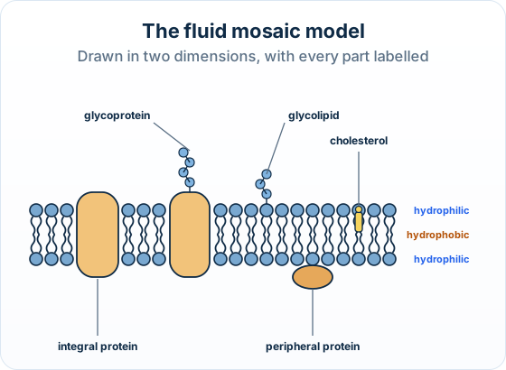
Common slips: Carbohydrate chains drawn on the inside, or peripheral proteins drawn inside the core, lose marks. Use a ruler for label lines and keep them clear of the drawing.
The big idea: Fluid: the parts drift sideways. Mosaic: many different molecules are scattered through the bilayer, like tiles.
The points to remember
- Fluid: phospholipids and many proteins are not fixed; they move sideways within the bilayer.
- Mosaic: proteins, glycoproteins, glycolipids and cholesterol are embedded throughout the bilayer; name them, not just 'molecules'.
- Hydrophilic regions: the phosphate heads and the carbohydrate chains, facing water.
- Hydrophobic region: the fatty acid tails of the core.
- Cholesterol sits among the tails and makes the membrane less permeable to small molecules.
How the model was tested: Freeze-fracture electron micrographs split membranes down the middle and showed proteins passing through the core. In 1972 Singer and Nicolson proposed the fluid mosaic model.
Feeling unprepared for exams?
Get a clear study plan, practice with real questions, and know exactly where you stand before exam day. No more guessing.
How it is asked: Paper 2 asks you to draw the model or to explain the words fluid and mosaic. Paper 1A shows a drawn model and asks which regions are hydrophilic, or which part does a job.
Draw a labelled diagram of the fluid mosaic model of a membrane.
Model answer plan
See the answer broken into clear points, with guidance on how each point earns marks.
And a one-mark Outline:
Outline why the structure of a membrane is described as a mosaic.
Model answer plan
See the answer broken into clear points, with guidance on how each point earns marks.