The big idea: The backbone of a chain has the same C=O and N-H groups again and again. Hydrogen bonds between them, at regular positions, coil the chain into helices or pleat it into sheets.
The points to remember
- Secondary structure is the coiling or pleating of parts of a chain.
- The backbone has a C=O and an N-H for every amino acid, at regular positions.
- Alpha helix: the chain coils, and each C=O bonds to the N-H four amino acids further along.
- Beta-pleated sheet: sections of chain lie side by side, with C=O on one bonding to N-H on the next.
- These are hydrogen bonds: each is weak, but many in regular positions make the structure stable.
Remember it as: Helix: coil, bonds up the coil. Sheet: pleats, bonds across.
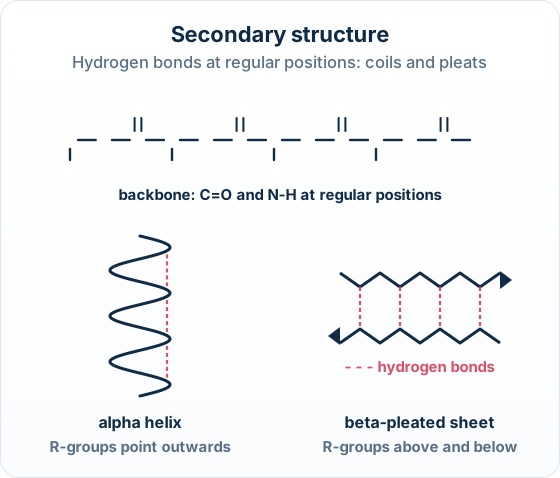
Backbone, not R-groups: The hydrogen bonds of secondary structure are between C=O and N-H of the backbone. R-groups stick out and are not part of them; R-group bonds hold the tertiary structure (2.2.9).
Free preview
This is the free notes preview
You're reading the free notes. Aimnova Pro unlocks the full study experience — and you can try it with your first topic free to keep:
- FlashcardsLock in vocabulary and key terms with spaced repetition.
- Practice questionsAnswer exam-style questions and get instant AI marking.
- Mock exams & past-paper vaultSit full mocks and see exactly how examiners award marks.
- Personalised study planA daily plan built around your exam date and weak areas.
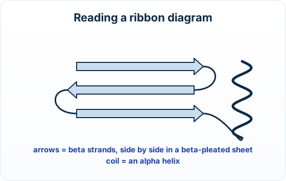
The big idea: Biologists draw proteins as ribbons: arrows for beta strands and coils for alpha helices, so you can see the secondary structure at a glance.
The points to remember
- In a ribbon diagram, a flat arrow is one beta strand; arrows side by side form a beta-pleated sheet.
- A coil or spiral is an alpha helix.
- Thin lines are loops that link helices and strands.
- Most proteins have both: helices and sheets, joined by loops, then folded further.
Real example: titin: Titin, the spring-like protein in muscle, is built from hundreds of repeating domains. Many of them are made almost entirely of beta-pleated sheets.
Practice with exam-style questions
Answer exam-style questions and get AI feedback that shows you exactly what examiners want to see in a full-marks response.
How it is asked: A ribbon diagram of part of a protein, and a two-mark Describe on the structures its arrows or coils show.
A ribbon diagram of one domain of a muscle protein shows several flat arrows lying side by side. Describe the secondary structures represented by the arrows.
Model answer plan
See the answer broken into clear points, with guidance on how each point earns marks.
And the same task for a coil:
Describe the secondary structure represented by the coils in a ribbon diagram of a protein.
Model answer plan
See the answer broken into clear points, with guidance on how each point earns marks.