Micrographs at Higher Level: The same statement as SL. HL papers give an electron micrograph and ask you to name the organelle labelled X, or to use only what you can see to decide the type of cell.
Practise this as you read
- State the name of a labelled organelle in full.
- Deduce the cell type with evidence you can see.
Free preview
This is the free notes preview
You're reading the free notes. Aimnova Pro unlocks the full study experience — and you can try it with your first topic free to keep:
- FlashcardsLock in vocabulary and key terms with spaced repetition.
- Practice questionsAnswer exam-style questions and get instant AI marking.
- Mock exams & past-paper vaultSit full mocks and see exactly how examiners award marks.
- Personalised study planA daily plan built around your exam date and weak areas.
The big idea: A micrograph is just a photograph taken down a microscope. You are not recalling a memorised picture — you are looking for evidence.
Read the clues and decide two things
- What type of cell is this? (prokaryotic or eukaryotic; plant, animal or fungus)
- Which structures can I see and name?
- Micrograph
- A photograph of a specimen taken through a microscope.
- Electron micrograph
- A micrograph taken with an electron microscope — high magnification, so small organelles such as mitochondria are visible.
- Organelle
- A structure inside a cell that does a specific job (for example the nucleus or a mitochondrion).
- Prokaryotic cell
- A cell with no nucleus and no membrane-bound organelles (for example a bacterium).
- Eukaryotic cell
- A cell with a nucleus and membrane-bound organelles (for example an animal, plant or fungal cell).
Two questions, every time: Train yourself to ask, in order:
1. Is there a nucleus? (the biggest clue to prokaryote vs eukaryote) 2. What else can I name? (cell wall, chloroplast, mitochondrion, vacuole…)
Stop wasting time on topics you know
Our AI identifies your weak areas and focuses your study time where it matters. No more overstudying easy topics.
The single most useful clue is the nucleus. Prokaryotic cells also differ in size and organelles.
Eukaryotic cell
- Has a nucleus — a large, dark, membrane-bound structure
Prokaryotic cell
- Has no nucleus: its DNA sits free in the cytoplasm as a single loop
- Much smaller
- No membrane-bound organelles at all (no mitochondria, no endoplasmic reticulum)
| Clue in the micrograph | Prokaryotic | Eukaryotic |
|---|---|---|
| A nucleus (dark, membrane-bound) | absent | present |
| Membrane-bound organelles (mitochondria, ER) | absent | present |
| DNA | free in the cytoplasm (a single loop) | inside a nucleus |
| Typical size | small (about 1–5 μm) | larger (about 10–100 μm) |
| A cell wall | always present | plant/fungus yes, animal no |
Once you know it is eukaryotic, a few extra clues tell you which kind:
Looks like a PLANT cell
- Has a cell wall (a straight, rigid edge)
- Often has chloroplasts (where photosynthesis happens)
- Has one large central vacuole
Looks like an ANIMAL cell
- No cell wall (a soft, irregular edge)
- No chloroplasts
- Only small vacuoles, if any
A fungus is a third option: A fungal cell is eukaryotic and has a cell wall (like a plant) but has no chloroplasts (like an animal). Its wall is made of chitin, not cellulose.
So: cell wall + chloroplasts → plant; cell wall + no chloroplasts → fungus; no cell wall → animal.
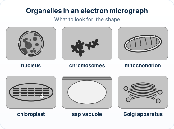
Naming organelles by how they LOOK: The exam also asks you to Identify a single labelled organelle — for example 'Identify organelle X' — so you must recognise the common organelles by their appearance in an electron micrograph, not just decide the cell type.
Each organelle has a shape you can spot. Match the picture to the name using the table below.
| Organelle | How it looks in the micrograph | What it does |
|---|---|---|
| Nucleus | Large and round, with a double membrane pierced by pores; dark chromatin inside | Stores the DNA |
| Mitochondrion | Oval (sausage-shaped) with folded inner membranes called cristae | Releases energy (respiration) |
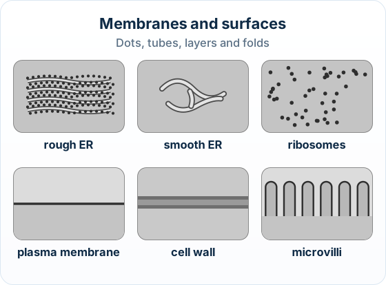
| Rough endoplasmic reticulum (rough ER) | Stacked membrane sheets studded with dotted ribosomes on the surface | Makes and transports proteins |
| Smooth endoplasmic reticulum (smooth ER) | A network of tubes with a smooth surface — no ribosomes (no dots) | Makes lipids; no protein synthesis |
| Golgi apparatus | A neat stack of flattened, curved sacs, often with small bubbles (vesicles) nearby | Modifies and packages proteins |
- Cristae
- The folds of the inner membrane of a mitochondrion — they give it its oval, ridged look.
- Ribosome
- A tiny dot-like structure where proteins are made; ribosomes stuck to membranes make the ER look 'rough'.
- Endoplasmic reticulum (ER)
- A system of membranes inside the cell. Rough ER carries ribosomes (dots); smooth ER does not.
- Golgi apparatus
- A stack of flattened membrane sacs that modifies and packages proteins ready to leave the cell.
ROUGH ER
- Membranes covered in dots (ribosomes)
- Often stacked, parallel sheets
- Job: makes and ships proteins
SMOOTH ER
- Membranes with a plain surface — no dots
- Looks like a network of curved tubes
- Job: makes lipids (not proteins)
The dots are the giveaway: The fastest way to separate the two kinds of ER is the ribosomes: dots on the membrane → rough ER; no dots → smooth ER.
And don't mix up the look-alikes — a mitochondrion is oval with internal cristae, whereas the Golgi apparatus is a stack of flat curved sacs.
The big idea: In an electron micrograph every structure has a shape you can learn. Match the shape to the name.
The points to remember
- Nucleus: the largest round shape, a double membrane with pores. Chromosomes show as thick dark threads in a dividing cell.
- Mitochondrion: an oval with inner folds (cristae). Chloroplast: larger, with stacks of membranes.
- Rough ER: membranes dotted with ribosomes. Smooth ER: tubes with no dots. Golgi: a stack of curved sacs.
- Sap vacuole: a large pale space. Cell wall: a thick layer outside the plasma membrane, a thin dark line.
- Microvilli: finger-like folds of the plasma membrane on some animal cells.
- A prokaryote has a pale nucleoid region of DNA, no nucleus, small ribosomes and a cell wall.
Remember it as: Dots on a membrane mean ribosomes: that is rough ER.
A prokaryote has none of these membrane-bound organelles. Look for these instead:
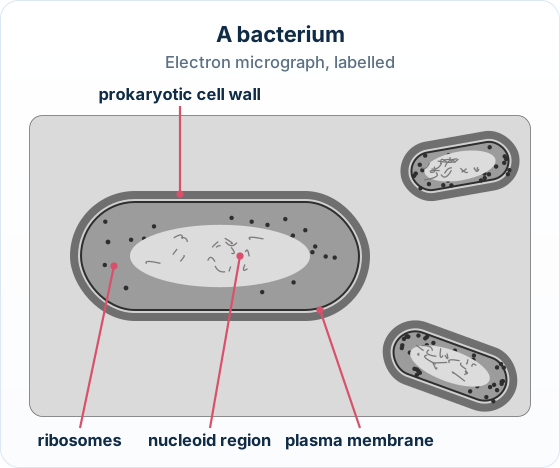
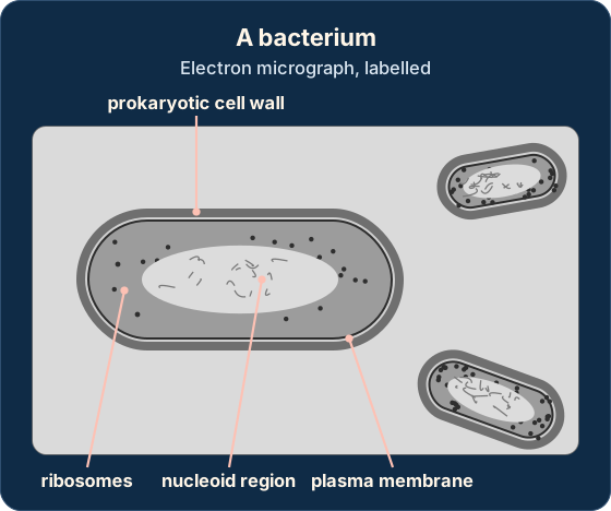
Name it in full: Write 'plasma membrane', not 'membrane' alone, and 'rough ER', not just 'ER'. A part name is not enough.
Chromosomes only show when a cell is dividing; the rest of the time the DNA is spread out as chromatin inside the nucleus.
Real example: Bacillus subtilis: Bacillus subtilis, a rod-shaped soil bacterium, has a thick cell wall that shows as a dark band all the way round it, outside the thin plasma membrane.
See how examiners mark answers
Answer exam-style questions with model answers. Learn exactly what earns marks and what doesn't.
The big idea: Read a micrograph in two steps: decide the type of cell from what you can see, then name its structures.
The points to remember
- Prokaryote: no nucleus, a nucleoid, small (about 1–5 µm). Eukaryote: a nucleus and membrane-bound organelles.
- Plant cell: a cell wall, chloroplasts and a large sap vacuole. Animal cell: no wall; it may have microvilli.
- A light micrograph shows the cell wall, nucleus, chloroplasts and vacuole. Smaller organelles need an electron micrograph.
- Give a reason you can see: a nucleus, a vacuole, chloroplasts. Never one you cannot, such as 80S ribosomes.
- A cell wall alone does not prove a eukaryote: bacteria have walls too.
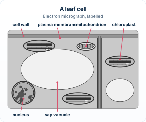
Real example: a spinach leaf: A palisade cell from a spinach leaf has a cellulose wall, many chloroplasts and a large sap vacuole that pushes them to the edges: a plant cell at a glance.
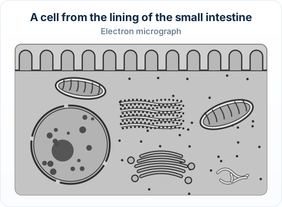
'Organelles' is not evidence on its own: Name one you can see, such as the nucleus or a mitochondrion. 'It has organelles' does not earn the mark.
How this is tested: You will be asked to read micrographs: deduce the cell type or identify a labelled structure.
Where it comes up
- Paper 2: you are often given a micrograph and asked to Deduce the cell type — and you must justify it with a feature you can see (for example 'no nucleus, so prokaryotic').
- Paper 1A: Identify a labelled organelle.
- Paper 1B: name a structure labelled X, or say from what you can see whether a cell is prokaryotic or eukaryotic.
Exam-style question — (a) deduce the cell type
An electron micrograph shows a small cell, about 3 μm across, with its DNA lying free in the cytoplasm and no nucleus or mitochondria visible.
Deduce whether this is a prokaryotic or a eukaryotic cell, giving a reason. [2]
How to score both marks
- Pick out the evidence. There is no nucleus and no membrane-bound organelles, and the DNA is free in the cytoplasm — these are all prokaryotic features.
- Answer the command term (Deduce). State the cell type and link it to the evidence: this is a prokaryotic cell because it has no nucleus (its DNA is free in the cytoplasm).
Final answer
Prokaryotic — because there is no nucleus (the DNA lies free in the cytoplasm) and no membrane-bound organelles.
An electron micrograph shows a cell from the lining of a human airway, with a scale bar of 2 µm. A large round structure with a double membrane and several ovals with inner folds can be seen. Deduce, using evidence from the micrograph, whether the cell is prokaryotic or eukaryotic.
Model answer plan
See the answer broken into clear points, with guidance on how each point earns marks.