The big idea: Blood is carried around the body in three kinds of vessel: arteries, veins and capillaries. Each one has a wall built to suit its job.
Arteries
- Carry blood away from the heart
- At high pressure
Veins
- Return blood to the heart
- At low pressure
Capillaries
- Where substances are exchanged with the tissues
Structure → function: The shape of the wall always matches the work the vessel has to do. This structure → function link is the whole point of this topic.
The three vessels in cross-section: the artery has a thick muscular and elastic wall with a narrow lumen (high pressure); the vein has a thin wall, a wide lumen and valves (low pressure); the capillary wall is just one cell thick for fast exchange.
Interactive diagram
Explore the labelled diagram, charts and maps for this topic in full study mode.
- Artery
- A blood vessel that carries blood away from the heart. It has a thick, muscular and elastic wall and a narrow lumen to withstand high pressure.
- Vein
- A blood vessel that returns blood to the heart. It has a thin wall, a wide lumen and valves to stop the low-pressure blood flowing backwards.
- Capillary
- A tiny blood vessel whose wall is only one cell thick. It is the site where substances are exchanged between the blood and the tissues.
- Lumen
- The hollow space inside a blood vessel through which the blood flows.
- Valve
- A flap inside a vein that opens to let blood pass towards the heart and closes to stop it flowing backwards.
A → away, capillaries → exchange: Arteries carry blood Away from the heart; veins bring it back.
Capillaries are the only vessels with a wall one cell thick, so they are the only place where exchange with the tissues can happen.
Free preview
This is the free notes preview
You're reading the free notes. Aimnova Pro unlocks the full study experience — and you can try it with your first topic free to keep:
- FlashcardsLock in vocabulary and key terms with spaced repetition.
- Practice questionsAnswer exam-style questions and get instant AI marking.
- Mock exams & past-paper vaultSit full mocks and see exactly how examiners award marks.
- Personalised study planA daily plan built around your exam date and weak areas.
Every feature of a vessel's wall is there for a reason. The trick is to say the structure and then the function it allows — never one without the other.
Work through each vessel by asking: what pressure does it carry, and what does its job need?
Artery — thick wall for high pressure: Blood leaves the heart in a powerful surge, so arteries carry blood at high pressure.
Their wall is thick, muscular and elastic:
the muscle and elastic tissue let the wall stretch as the surge passes and then recoil, which withstands the high pressure and helps push the blood onward.
The narrow lumen keeps the pressure high all the way along.
Vein — thin wall and valves for low pressure: By the time blood returns, most of the pressure is gone, so veins carry blood at low pressure.
Vein structure → function
- A thick wall is not needed, so the wall is thin
- The lumen is wide (less resistance to the slow-moving blood)
- Because the pressure is so low, blood could slip backwards
- So veins have valves that close to stop backflow and keep blood moving towards the heart
Capillary — one cell thick for exchange: Capillaries are where the blood actually does its job: exchanging oxygen, glucose, carbon dioxide and wastes with the tissues.
Capillary structure → function
- The wall is only one cell thick, giving a short diffusion distance, so substances cross quickly
- They are very narrow (a red blood cell almost fills the lumen)
- They form a huge branching network, giving a large surface area close to every cell
Compare the walls: thick in the artery to withstand high pressure, thin in the vein, and only one cell thick in the capillary so substances can diffuse across quickly.
Interactive diagram
Explore the labelled diagram, charts and maps for this topic in full study mode.
Artery
- Carries blood away from the heart
- Thick muscular and elastic wall
- Narrow lumen
- High pressure — wall stretches and recoils
- No valves
Vein
- Returns blood to the heart
- Thin wall
- Wide lumen
- Low pressure
- Valves stop backflow
Match the wall to the pressure: Thick wall = high pressure (artery). Thin wall + valves = low pressure (vein). One cell thick = exchange (capillary).
If you can match each wall to the pressure or job it handles, you can answer almost any question on this micro.
| Feature | Artery | Vein | Capillary |
|---|---|---|---|
| Direction of blood | Away from the heart | Back to the heart | Through the tissues |
| Wall | Thick — muscular and elastic | Thin | One cell thick |
| Lumen (space inside) | Narrow | Wide | Very narrow (one cell wide) |
| Pressure of blood | High | Low | Falling (low) |
| Valves? | No | Yes — stop backflow | No |
| Main job | Carry blood at high pressure | Return blood at low pressure | Exchange materials with tissues |
Learn what examiners really want
See exactly what to write to score full marks. Our AI shows you model answers and the key phrases examiners look for.
The big idea: In a micrograph, compare the wall with the lumen. A thick wall round a narrow lumen is an artery; a thin wall round a wide lumen is a vein.
The points to remember
- Artery: wall about as thick as the lumen is wide; thick layer of smooth muscle and elastic fibres.
- Vein: thin wall next to a wide lumen; little muscle and elastic tissue.
- Both: a tough outer layer of collagen and a smooth lining of endothelium, one cell thick.
- An artery stays round when cut; a vein is often flattened or collapsed.
- Only veins have valves; they show when a vein is cut along its length.
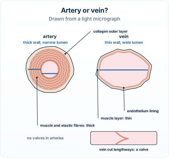
Remember it as: Artery: thick wall, small hole. Vein: thin wall, big hole.
Real example: the wrist. The radial artery at the wrist keeps its round shape and pulses under your finger; the cephalic vein beside it is soft and flattens when you press it.
Two traps: Judge the wall relative to the lumen, not by the size of the whole vessel: a small artery can be narrower overall than a large vein.
When you label an artery, the line must touch the artery, not the space between vessels.
How this is tested: Questions ask you to compare vessel walls, identify vessels in a micrograph, or outline how capillaries suit exchange.
What the questions ask
- A favourite task: distinguish the wall structure of an artery from that of a vein
- For 2 marks you need paired contrasts (artery thick / vein thin; artery no valves / vein has valves)
- You may be shown a micrograph of two vessels and asked to identify the artery and justify it from its structure (thick wall, narrow lumen)
- Capillary questions usually ask you to describe or outline how they are adapted for exchange (one-cell-thick wall → short diffusion distance; large surface area)
Exam-style question — distinguish artery and vein walls
Distinguish between the wall structure of an artery and that of a vein. [2]
How to score both marks
- Contrast the wall thickness. An artery has a thick wall, with a lot of muscle and elastic tissue, whereas a vein has a thin wall.
- Contrast a second feature. An artery has a narrow lumen and no valves, whereas a vein has a wide lumen and valves to stop backflow.
Final answer
An artery has a thick, muscular wall with a narrow lumen and no valves, whereas a vein has a thin wall with a wide lumen and valves to prevent backflow.
Why this scores full marks: Each mark is a paired contrast — artery does X whereas vein does Y — not two facts about arteries on their own.
On a Distinguish question, the word 'whereas' (or 'but') is what earns the mark: describing only one vessel gets zero.
| Wall feature | Artery | Vein |
|---|---|---|
| Thickness | Thick wall | Thin wall |
| Muscle and elastic tissue | Lots (thick muscular and elastic layer) | Little |
| Lumen | Narrow | Wide |
| Valves along the vessel | Absent | Present (stop backflow) |
A 7-mark Explain, in the papers' style:
Explain how the structures of arteries and veins are adapted to their functions.
Model answer plan
See the answer broken into clear points, with guidance on how each point earns marks.