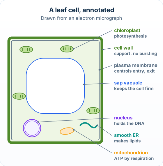
The big idea: A drawing from an electron micrograph shows only what you can see, in clear lines, with each part labelled and its job written beside it.
The points to remember
- Use clear, single lines: no shading and no colouring in.
- Keep the proportions: a mitochondrion is smaller than the nucleus; draw what the micrograph shows.
- Put labels at the side, with ruled lines that touch the structure and never cross each other.
- To annotate, add the function beside each label: 'Golgi apparatus: packs proteins'.
- A Draw question gives about one mark per structure drawn correctly and labelled.
Remember it as: Draw it, label it, say what it does.
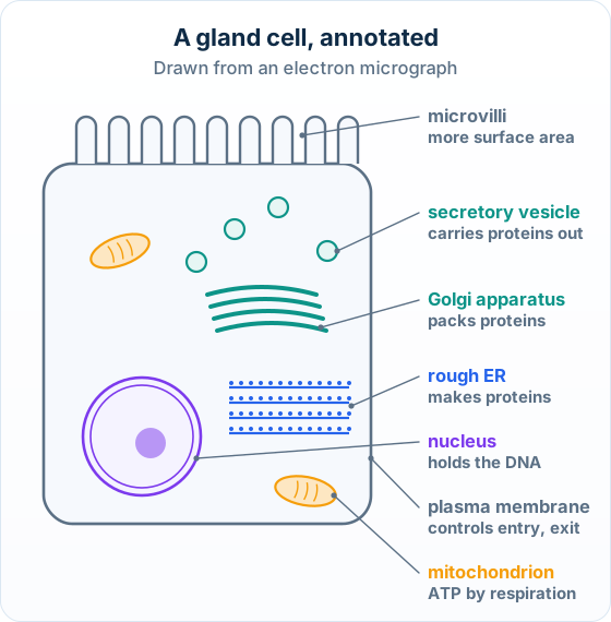
Real example: a pancreas cell: Cells in your pancreas make digestive enzymes. An electron micrograph shows them packed with rough ER, Golgi stacks and secretory vesicles, the route every enzyme takes out of the cell.
Lines and labels earn the marks; colour and shading do not.
Only what you can see: Label only structures the micrograph shows. A label for something hidden, such as DNA inside a mitochondrion, does not score.
Free preview
This is the free notes preview
You're reading the free notes. Aimnova Pro unlocks the full study experience — and you can try it with your first topic free to keep:
- FlashcardsLock in vocabulary and key terms with spaced repetition.
- Practice questionsAnswer exam-style questions and get instant AI marking.
- Mock exams & past-paper vaultSit full mocks and see exactly how examiners award marks.
- Personalised study planA daily plan built around your exam date and weak areas.
The big idea: The nucleus is drawn as two close circles with gaps (pores) through both, chromatin and a nucleolus inside.
The points to remember
- Nuclear envelope: two membranes, drawn as two close lines; it separates the DNA from the cytoplasm.
- Nuclear pores: gaps through both membranes; mRNA leaves the nucleus through them.
- Chromatin: the DNA with its proteins, drawn as darker patches.
- Nucleolus: a dense round area where ribosomes are made.
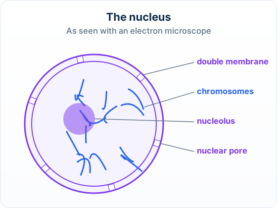
Draw a labelled diagram of a nucleus from a eukaryotic cell, as seen using an electron microscope.
Model answer plan
See the answer broken into clear points, with guidance on how each point earns marks.
How to draw the nucleus for full marks: a DOUBLE-membrane nuclear envelope (two lines) pierced by nuclear pores, with the chromatin (DNA) and a dense round nucleolus inside.
Interactive diagram
Explore the labelled diagram, charts and maps for this topic in full study mode.
Check your drawing: A good electron-micrograph nucleus has four labelled features: the double membrane (nuclear envelope), the nuclear pores, the chromatin and the nucleolus. Drawing the envelope as a single line is the most common lost mark.
Feeling unprepared for exams?
Get a clear study plan, practice with real questions, and know exactly where you stand before exam day. No more guessing.
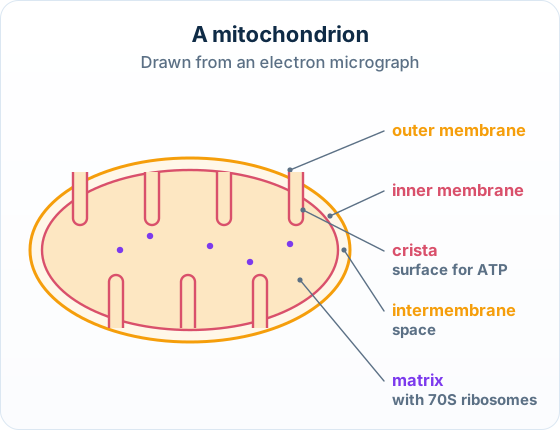
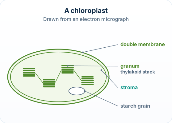
The big idea: Both organelles have two membranes. Inside, a mitochondrion has folds (cristae); a chloroplast has stacks (grana).
The points to remember
- Mitochondrion: outer membrane, inner membrane folded into cristae, the intermembrane space, the matrix.
- Annotate the cristae: a large surface for the last stage of aerobic respiration, which makes ATP.
- Chloroplast: double membrane, grana (stacks of thylakoids) where light is absorbed, stroma, starch grains.
- Small dots in the matrix or stroma are 70S ribosomes.
A chloroplast is drawn the same way, with stacks instead of folds:
Two membranes, two lines: Draw the outer and inner membranes as two separate lines. One line loses the mark for the membranes.
The big idea: Every other structure has a simple shape to draw and one job to write beside it.
| Structure | Draw it as | Annotate with |
|---|---|---|
| Sap vacuole | a large space with a single line round it | stores sap; keeps the cell firm |
| Golgi apparatus | a stack of curved sacs, vesicles at the ends | modifies and packs proteins |
| Rough ER | parallel lines with dots (ribosomes) | makes proteins for export |
| Smooth ER | curved tubes, no dots | makes lipids |
| Chromosomes | thick dark threads (dividing cell) | carry the genes (DNA) |
| Cell wall | a thick layer outside the membrane | support; stops the cell bursting |
| Plasma membrane | one thin line round the cytoplasm | controls what enters and leaves |
| Secretory vesicles | small circles near the membrane | carry proteins out of the cell |
| Microvilli | finger-like folds on one surface | more surface area for absorption |
Remember it as: Rough ER makes, Golgi packs, vesicles carry out.
Secretory vesicles in the drawing: Draw them as small circles between the Golgi apparatus and the plasma membrane, on the side where the cell releases its product.
Memorize terms 3x faster
Smart flashcards show you cards right before you forget them. Perfect for definitions and key concepts.
How it is tested: Paper 1B asks you to draw and label an organelle seen in an electron micrograph, for three marks.
An electron micrograph shows part of a heart muscle cell. Draw a labelled diagram of a mitochondrion as seen in the micrograph.
Model answer plan
See the answer broken into clear points, with guidance on how each point earns marks.
Marks for labels: Each structure must be drawn and labelled: a label on a structure that is not drawn does not score.