Neurotransmitter release at Higher Level: Shared with SL; at HL drugs and pesticides that act on synapses follow in 3.5.12.
Practise this as you read
- Describe neurotransmitter release.
- Explain the role of calcium.
Free preview
This is the free notes preview
You're reading the free notes. Aimnova Pro unlocks the full study experience — and you can try it with your first topic free to keep:
- FlashcardsLock in vocabulary and key terms with spaced repetition.
- Practice questionsAnswer exam-style questions and get instant AI marking.
- Mock exams & past-paper vaultSit full mocks and see exactly how examiners award marks.
- Personalised study planA daily plan built around your exam date and weak areas.
Synaptic transmission is a chain of cause and effect. Each step makes the next one happen, so it is easiest to learn as an ordered sequence.
Split it into two halves
- First the presynaptic neuron releases the neurotransmitter
- Then the postsynaptic neuron responds to it
Step 1 — release at the presynaptic membrane: When the impulse reaches the presynaptic neuron's terminal:
1. Calcium ions (Ca²⁺) enter the presynaptic neuron through channels that open.
2. This makes synaptic vesicles fuse with the presynaptic membrane.
3. The vesicles release neurotransmitter into the synaptic cleft by exocytosis.
Release step: vesicles in the presynaptic neuron fuse with the membrane and empty neurotransmitter (amber dots) into the synaptic cleft by exocytosis.
Interactive diagram
Explore the labelled diagram, charts and maps for this topic in full study mode.
Step 2 — response at the postsynaptic membrane
Diffuses
The neurotransmitter then diffuses across the cleft.
Binds
It binds to receptors on the postsynaptic membrane.
Na⁺ enters
Binding opens ion channels, so sodium ions (Na⁺) enter the postsynaptic neuron.
Depolarises
The postsynaptic membrane depolarises — this is an excitatory postsynaptic potential (EPSP).
New impulse
If the depolarisation is big enough to reach threshold, a new nerve impulse fires in the postsynaptic neuron.
Diffuse → bind → Na⁺ in → EPSP → new impulse
Receiving step: neurotransmitter crosses the narrow cleft and binds receptors on the postsynaptic membrane, opening ion channels and depolarising it toward a new impulse.
Interactive diagram
Explore the labelled diagram, charts and maps for this topic in full study mode.
- Exocytosis
- The process in which vesicles fuse with the cell membrane and empty their contents to the outside — how neurotransmitter is released.
- Excitatory postsynaptic potential (EPSP)
- A depolarisation of the postsynaptic membrane caused by neurotransmitter binding — it makes a new impulse MORE likely.
- Threshold
- The level of depolarisation that must be reached for a new nerve impulse (action potential) to fire.
The whole sequence in order: Impulse arrives → Ca²⁺ enters → vesicles fuse → neurotransmitter released (exocytosis) → diffuses across cleft → binds receptors → Na⁺ enters → membrane depolarises (EPSP) → new impulse fires.
Write the steps in this order when you describe the process.
| Step | What happens | Result |
|---|---|---|
| 1. Impulse arrives | A nerve impulse reaches the presynaptic neuron's terminal | The terminal is depolarised |
| 2. Calcium enters | Calcium ions (Ca²⁺) flow into the presynaptic neuron | Vesicles are triggered to move to the membrane |
| 3. Vesicles fuse | Synaptic vesicles fuse with the presynaptic membrane | Neurotransmitter is released by exocytosis |
| 4. Diffusion | Neurotransmitter diffuses across the synaptic cleft | It reaches the postsynaptic membrane |
| 5. Binding | Neurotransmitter binds receptors on the postsynaptic membrane | Ion channels open and Na⁺ enters |
| 6. New impulse | The postsynaptic membrane depolarises; if threshold is reached | A new nerve impulse is triggered |
Why the gap is so narrow: The synaptic cleft is extremely thin.
That matters because the neurotransmitter crosses it by diffusion, and diffusion is fast only over a short distance.
A narrow gap therefore means a short diffusion distance, so the signal is passed across quickly — a favourite 'Suggest an advantage' answer.
Presynaptic side
- Holds synaptic vesicles full of neurotransmitter
- Ca²⁺ enters when the impulse arrives
- Vesicles fuse and release neurotransmitter (exocytosis)
- The sender — signal leaves here
Postsynaptic side
- Carries receptor proteins
- Neurotransmitter binds the receptors
- Na⁺ enters, the membrane depolarises (EPSP)
- The receiver — a new impulse starts here
Memorize terms 3x faster
Smart flashcards show you cards right before you forget them. Perfect for definitions and key concepts.
The big idea: When an action potential depolarizes the presynaptic membrane, calcium channels open and calcium ions diffuse in. Inside the neuron, calcium acts as a signalling chemical: it makes vesicles fuse with the membrane and release neurotransmitter by exocytosis.
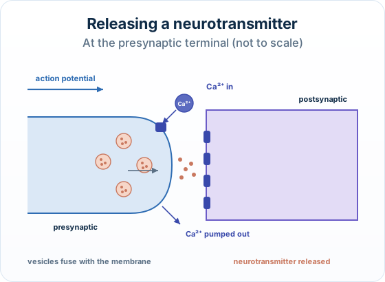
The points to remember
- Action potential depolarizes the presynaptic terminal.
- Calcium channels open: Ca²⁺ diffuses in.
- Ca²⁺ is the signal inside the neuron.
- Vesicles fuse with the membrane: exocytosis.
- Ca²⁺ is pumped out, so release stops.
Remember it as: Impulse, calcium in, vesicles out.
Real example: botulinum toxin. The toxin used in Botox stops vesicles at neuromuscular junctions from releasing acetylcholine, so the muscle stays relaxed.
Trap: It is calcium, not sodium, that triggers the vesicles to fuse.
How this is tested: Two Describe questions come up often, and data questions may show a synapse micrograph.
Describe: release at the presynaptic membrane
- How a neurotransmitter is released at the presynaptic membrane
- Score it as an ordered chain: Ca²⁺ in → vesicles fuse → exocytosis into the cleft
Describe: generating an EPSP
- How an excitatory postsynaptic potential (EPSP) is generated
- Neurotransmitter binds receptors → ion channels open → Na⁺ in → depolarisation
With a synapse micrograph, you may have to
- Deduce what the vesicles hold
- Distinguish the presynaptic from the postsynaptic membrane
- Suggest why a narrow cleft is an advantage
Exam-style question — describe neurotransmitter release
Describe how a neurotransmitter is released at the presynaptic membrane when a nerve impulse arrives. [3]
Step by step
- Calcium enters. The arriving impulse opens channels, so calcium ions (Ca²⁺) enter the presynaptic neuron.
- Vesicles fuse. The calcium causes synaptic vesicles (which hold the neurotransmitter) to move to and fuse with the presynaptic membrane.
- Exocytosis. The vesicles release the neurotransmitter into the synaptic cleft by exocytosis.
Final answer
The impulse causes calcium ions (Ca²⁺) to enter the presynaptic neuron; this makes synaptic vesicles fuse with the presynaptic membrane; they release neurotransmitter into the synaptic cleft by exocytosis.
Why this works: Each sentence is a separate, ordered event — calcium in, vesicles fuse, exocytosis.
A 'Describe' of a process wants the cause→effect chain in order, not one idea written three ways. Name Ca²⁺ and exocytosis.
| Feature | Presynaptic membrane | Postsynaptic membrane |
|---|---|---|
| Which neuron | The neuron the impulse arrives at FIRST (the sender) | The neuron the signal passes TO (the receiver) |
| What it holds | Synaptic vesicles full of neurotransmitter | Receptor proteins that the neurotransmitter binds to |
| Its job | RELEASES neurotransmitter into the cleft (exocytosis) | RECEIVES neurotransmitter; binding opens ion channels |
| Direction of signal | Signal leaves here | Signal continues from here |
Another question in the papers' style:
Explain the role of calcium ions in releasing a neurotransmitter.
Model answer plan
See the answer broken into clear points, with guidance on how each point earns marks.