The big idea: The heart is a muscular pump that pushes blood around the body. It is really two pumps side by side.
Four chambers
- Two thin-walled atria on top receive blood
- Two thicker-walled ventricles below pump it out
- A wall called the septum separates the two sides, so the two kinds of blood never mix
Right side
- Handles deoxygenated blood
- Sends it to the lungs
Left side
- Handles oxygenated blood
- Sends it to the body
The four chambers and four great vessels. Blue = deoxygenated blood (right side → lungs); red = oxygenated blood (left side → body). The left ventricle has the thickest wall because it pumps to the whole body.
Interactive diagram
Explore the labelled diagram, charts and maps for this topic in full study mode.
- Atrium (plural: atria)
- An upper, thin-walled chamber of the heart that receives blood arriving in the veins and passes it down into a ventricle.
- Ventricle
- A lower, thick-walled chamber of the heart that pumps blood out into an artery. The left ventricle has the thickest wall.
- Septum
- The muscular wall that separates the right side of the heart from the left, keeping deoxygenated and oxygenated blood apart.
- Atrioventricular (AV) valve
- A valve between an atrium and a ventricle that stops blood flowing backwards into the atrium when the ventricle contracts.
- Semilunar valve
- A valve at the exit of each ventricle (into the pulmonary artery and the aorta) that stops blood flowing back into the ventricle.
- Deoxygenated / oxygenated blood
- Deoxygenated blood is low in oxygen (returning from the body); oxygenated blood is high in oxygen (returning from the lungs).
Why the left ventricle has the thickest wall: The left ventricle pumps blood to the whole body, so it must generate a high pressure. That needs a lot of muscle, so its wall is the thickest of the four chambers.
Thinner walls elsewhere
- The right ventricle pumps only to the nearby lungs at lower pressure, so its wall is thinner
- The atria barely pump at all (just down into the ventricles), so their walls are thinnest
| Chamber | Blood it holds | Receives from / pumps to | Wall |
|---|---|---|---|
| Right atrium | deoxygenated | receives from the vena cava (body) | thin |
| Right ventricle | deoxygenated | pumps to the pulmonary artery (lungs) | thicker than atrium |
| Left atrium | oxygenated | receives from the pulmonary vein (lungs) | thin |
| Left ventricle | oxygenated | pumps to the aorta (whole body) | THICKEST — most muscle |
Free preview
This is the free notes preview
You're reading the free notes. Aimnova Pro unlocks the full study experience — and you can try it with your first topic free to keep:
- FlashcardsLock in vocabulary and key terms with spaced repetition.
- Practice questionsAnswer exam-style questions and get instant AI marking.
- Mock exams & past-paper vaultSit full mocks and see exactly how examiners award marks.
- Personalised study planA daily plan built around your exam date and weak areas.
The big idea: Every part of the heart helps push blood into the arteries at high pressure, in one direction only.
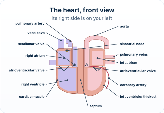
| Structure | How it is adapted |
|---|---|
| Cardiac muscle | Contracts strongly and rhythmically, without tiring |
| Pacemaker (sinoatrial node) | Starts each heartbeat, setting the heart rate |
| Atria | Thin walls: collect blood and push it a short way into the ventricles |
| Ventricles | Thick muscular walls: pump blood out at high pressure; the left is thickest |
| Atrioventricular valves | Shut when the ventricles contract: no backflow into the atria |
| Semilunar valves | Shut when the ventricles relax: no backflow from the arteries |
| Septum | Keeps oxygenated and deoxygenated blood apart |
| Coronary arteries | Supply the heart muscle itself with oxygen and glucose |
The points to remember
- One-way route: vena cava → right atrium → right ventricle → pulmonary artery (lungs) → pulmonary vein → left atrium → left ventricle → aorta.
- Valves open and shut with pressure, so blood can only move forwards.
- The left ventricle has the thickest wall because it pumps to the whole body.
Remember it as: Veins in, atria, ventricles, arteries out: valves keep it one way.
Real example: a heart transplant. When a donor heart is fitted, its own coronary arteries go with it: they are the heart muscle's only blood supply.
Two traps: The heart muscle is not fed by the blood inside its chambers: it needs the coronary arteries.
Vessels leaving the heart are arteries (aorta, pulmonary artery); vessels entering it are veins.
Memorize terms 3x faster
Smart flashcards show you cards right before you forget them. Perfect for definitions and key concepts.
How this is tested: Heart questions ask you to trace blood flow, read a pressure graph, or explain double circulation.
What the questions ask
- 2-mark Describe: trace the circulation of blood from the heart to the lungs
- Name the chambers and vessels in order, and use the word deoxygenated
- 1-mark data question: read a cardiac-cycle pressure graph and match the curves to the atrium, ventricle and aorta
- Or deduce the cause of a heart sound from where the lines cross
- 2-mark Explain: why blood must be pumped twice to get from the lungs to the body — the double-circulation reasoning
Exam-style question — trace blood from the heart to the lungs
Describe the circulation of deoxygenated blood from the heart to the lungs. [2]
How to score both marks
- Start where the deoxygenated blood arrives. Deoxygenated blood returns from the body in the vena cava and enters the right atrium, then passes into the right ventricle.
- Follow it out to the lungs. The right ventricle contracts and pumps the blood into the pulmonary artery, which carries it to the lungs to be oxygenated.
Final answer
Deoxygenated blood enters the right atrium, passes into the right ventricle, and is pumped through the pulmonary artery to the lungs.
Follow the blue side for the pathway to the lungs: vena cava → right atrium → right ventricle → pulmonary artery → lungs. Then the red side for the pathway to the body: pulmonary vein → left atrium → left ventricle → aorta → body.
Interactive diagram
Explore the labelled diagram, charts and maps for this topic in full study mode.
Why this scores full marks: The structures are named in the correct order and the route ends at the lungs.
A common slip is forgetting the pulmonary artery (the vessel that actually carries the blood out), or writing 'vein' instead — the vessel leaving the heart is always an artery.
Single and double circulation
- How a mammal's double circulation differs from a fish's single circulation is taught on 2.7.14.
A 3-mark Outline, in the papers' style:
Outline how structures in the heart and veins prevent the backflow of blood.
Model answer plan
See the answer broken into clear points, with guidance on how each point earns marks.