The big idea: Tissue fluid forms when blood pressure forces plasma out of capillaries, and most of it drains back in further along.
The points to remember
- Blood from an arteriole is at high hydrostatic pressure.
- At the arteriole end, this pressure forces plasma through the capillary wall: pressure filtration.
- Water and small solutes (glucose, oxygen, ions, amino acids) leave; plasma proteins and blood cells stay in.
- Pressure falls along the capillary; at the venule end it is lower, so most tissue fluid drains back in.
- The rest drains into lymph ducts (2.7.13).
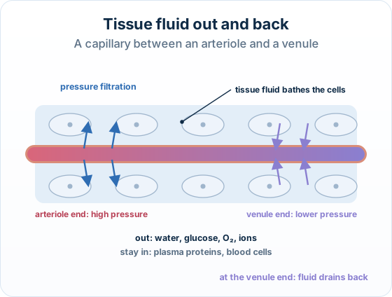
Arteriole end
- Blood pressure higher than in tissue fluid
- Plasma is forced out
- Tissue fluid forms
Venule end
- Blood pressure lower
- Fluid drains back in
- Tissue fluid is reabsorbed
Remember it as: Pushed out at the start, drains back at the end.
Real example: swollen ankles. After hours sitting still on a long flight, more fluid leaves the capillaries in the legs than drains back, so ankles swell with tissue fluid.
Two traps: Tissue fluid forms because of pressure, not because the capillary wall is 'more permeable' at one end.
Proteins do not leave: they are too large, so tissue fluid has almost no protein.
Free preview
This is the free notes preview
You're reading the free notes. Aimnova Pro unlocks the full study experience — and you can try it with your first topic free to keep:
- FlashcardsLock in vocabulary and key terms with spaced repetition.
- Practice questionsAnswer exam-style questions and get instant AI marking.
- Mock exams & past-paper vaultSit full mocks and see exactly how examiners award marks.
- Personalised study planA daily plan built around your exam date and weak areas.
Start Studying Free Full access to Aimnova Pro · cancel anytime
How it is asked: Expect a capillary-bed diagram with a region marked: what happens there, and why.
IB-style questionExplain[3 marks]
Explain how tissue fluid is formed and returned to the blood in a capillary bed.
Model answer plan
See the answer broken into clear points, with guidance on how each point earns marks.