Cell size at Higher Level: The same statement as SL. At HL the sizes lead on to surface area-to-volume ratios and to the adaptations of gametes, muscle cells and pneumocytes on the next pages.
Practise this as you read
- Compare the sizes of named human cells.
- Explain how a cell's size suits its function.
Free preview
This is the free notes preview
You're reading the free notes. Aimnova Pro unlocks the full study experience — and you can try it with your first topic free to keep:
- FlashcardsLock in vocabulary and key terms with spaced repetition.
- Practice questionsAnswer exam-style questions and get instant AI marking.
- Mock exams & past-paper vaultSit full mocks and see exactly how examiners award marks.
- Personalised study planA daily plan built around your exam date and weak areas.
The big idea: A specialized cell is a cell whose structure has been altered to do one particular job really well. The rule for this whole topic: structure follows function.
How it works
- In a multicellular organism, cells differentiate — they switch on different genes
- So each cell type ends up with a shape and contents that suit its function
- If you can see (or are told) a cell's feature, you can work out the job it does — and vice versa
Six specialized cells — each card names the cell, shows its shape, and gives the adaptation that suits its job. The shape always follows the function.
Interactive diagram
Explore the labelled diagram, charts and maps for this topic in full study mode.
- Specialized cell
- A cell with a structure adapted to carry out a particular function efficiently (for example a red blood cell adapted to carry oxygen).
- Differentiation
- The process by which an unspecialized cell becomes a particular specialized cell type, by expressing (switching on) a specific set of its genes.
- Adaptation
- A structural feature of a cell that makes its function easier or more efficient (for example microvilli that increase surface area).
- Structure–function relationship
- The idea that a cell's shape and contents (its structure) match the job it has to do (its function).
- Surface area to volume ratio
- How much surface a cell has compared with its volume. Features that increase surface area (folds, projections) speed up exchange and absorption.
How to read any specialized cell: For every specialized cell, ask two questions:
1. What is unusual about its structure? (its shape, what it contains, what it lacks)
2. How does that feature make its job easier?
Linking those two is exactly what the exam rewards — never describe a structure without saying the function it serves.
Study smarter, not longer
Most students waste 40% of study time on topics they already know. Our AI tracks your progress and optimizes every minute.
Each specialized cell has a feature that fits its job. The skill is to pair each structural feature with the function it makes possible — cause to effect.
Work through the table below one row at a time: name the feature, then state what it lets the cell do.
| Specialized cell | Structural feature (what you see) | Function it makes possible |
|---|---|---|
| Red blood cell | Biconcave disc shape; no nucleus; packed with haemoglobin | Large surface area and more room for haemoglobin → carries lots of oxygen |
| Intestine lining cell | Microvilli (folds) on its surface; many mitochondria | Large surface area and energy (ATP) → absorbs digested nutrients quickly |
| Sperm cell | Tail (flagellum); many mitochondria in the mid-piece | Energy and a tail → swims to reach and fertilise the egg |
| Neuron (nerve cell) | Very long fibre (axon) | Carries electrical impulses over long distances in the body |
| Root hair cell | Long, thin projection out into the soil | Large surface area → absorbs water and minerals efficiently |
| Palisade mesophyll cell | Column shape near the upper leaf surface; packed with chloroplasts | Catches the most light → carries out most of the leaf's photosynthesis |
| Cell | Feature | What it lets the cell do |
|---|---|---|
| Red blood cell | Biconcave and has no nucleus, so it is packed with haemoglobin and has a large surface area | Carries the most oxygen |
| Intestine lining cell | Covered in microvilli (tiny folds) and full of mitochondria | A huge surface area and plenty of energy to absorb nutrients quickly |
| Sperm cell | Has a tail and many mitochondria | Energy to swim to the egg |
Relative size matters too: Questions also test relative size. Most body cells are tiny (about 10–30 µm).
Largest
- The egg cell (ovum) is the largest human cell
- It stores food reserves for the early embryo
Among the smallest
- The sperm cell — stripped down to swim
- The red blood cell — small and flexible to fit through narrow capillaries
| Cell | Relative size | Why |
|---|---|---|
| Egg cell (ovum) | Largest — by far the biggest human cell | Stores food reserves (yolk / cytoplasm) for the early embryo before implantation |
| Most body cells | Tiny (about 10–30 µm across) | Small size keeps a large surface-area-to-volume ratio for exchange |
| Sperm cell | Among the smallest | Stripped down to a head and a tail so it can swim quickly |
| Red blood cell | Very small (about 7 µm) | Small and flexible to squeeze through narrow capillaries |
Animal cell adaptations
- Red blood cell — biconcave + no nucleus → carries O₂
- Intestine cell — microvilli + mitochondria → absorbs nutrients
- Sperm cell — tail + mitochondria → swims to the egg
- Neuron — long axon → carries impulses far
Plant cell adaptations
- Palisade cell — packed chloroplasts near the top → photosynthesis
- Root hair cell — long projection → absorbs water and minerals
- Both increase surface area or capture light
- Plant cells specialize for making food and taking up water
An easy way to remember: Shape suits the job. A swimmer gets a tail, an absorber gets folds or projections (more surface area), a light-catcher gets chloroplasts, and an oxygen-carrier drops its nucleus to make room.
Big cell that feeds an embryo = the egg; small cells that move = sperm and red blood cells.
The big idea: Human cells range from a few micrometres to about a metre: each cell's size suits its job.
The points to remember
- Red blood cells are tiny (about 7.5 µm), so they squeeze through narrow capillaries.
- White blood cells are a little larger (about 12 µm): many engulf pathogens.
- Sperm are small and streamlined, with little cytoplasm, so they can swim fast to the egg.
- The egg is the largest human cell (about 0.1 mm): its cytoplasm holds food for the early embryo.
- Neurons can be up to about 1 m long, to carry impulses from the spine to the feet.
- Striated muscle fibres are very long (up to about 10 cm), so one fibre can pull along much of a muscle's length.
Remember it as: Feed it, big egg; swim it, small sperm; squeeze it, small red cell; reach it, long neuron.
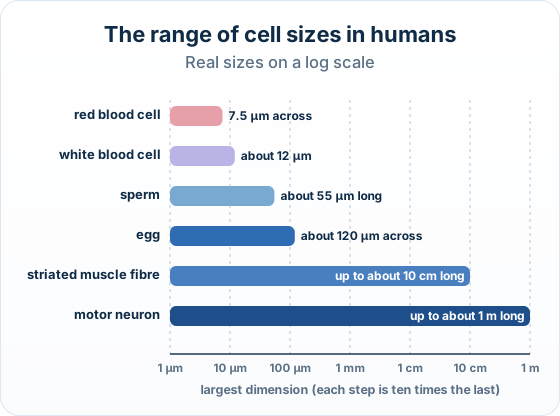
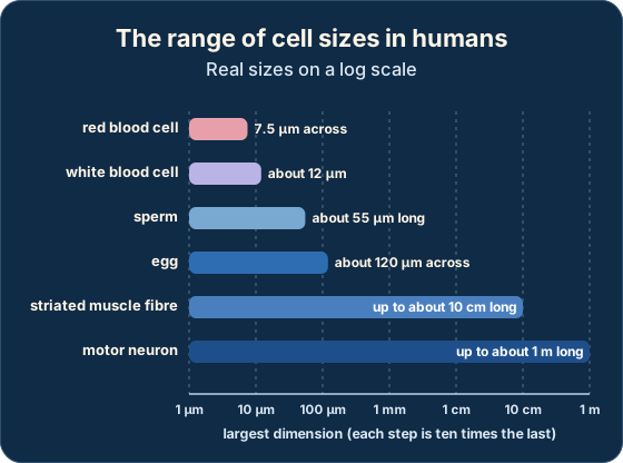
Real example: the longest human cells: Motor neurons that run from the base of the spine to the muscles of the big toe can be about 1 m long, yet their cell body is less than 0.1 mm across.
Practice with exam-style questions
Answer exam-style questions and get AI feedback that shows you exactly what examiners want to see in a full-marks response.
How this is tested: The classic question here is a 3-mark Explain: explain how a specialized cell's structure adapts it to its function.
What to expect
- You are shown (or told about) a specialized cell
- It is a data favourite on Paper 1B: an electron micrograph is given
- First name the labelled cell, then read the visible adaptations off the image and link each one to a function
How it is marked: The marking pattern is always feature → what it lets the cell do — one mark per correctly linked pair. A bare list of features with no function scores nothing.
Exam-style question — explain a cell's adaptations to its function
A cell from the lining of the small intestine has its surface folded into many microvilli and contains a large number of mitochondria. Explain how the structure of this cell adapts it to its function of absorbing digested food. [3]
How to score all three marks
- Microvilli → surface area. The microvilli (folds) greatly increase the cell's surface area, so more nutrients can be absorbed at the same time.
- Mitochondria → energy. The many mitochondria release energy (ATP) by respiration, which powers the active transport of nutrients into the cell.
- Link to the function. Together these features let the cell absorb digested food quickly and efficiently from the gut. (Award 1 mark for each correctly linked structure→function pair, max 3.)
Final answer
Microvilli increase the surface area for absorption; many mitochondria supply the energy (ATP) for active transport of nutrients — so the cell absorbs digested food quickly and efficiently.
Why this scores full marks: Each mark is a pair: a structure named and the function it serves — microvilli for surface area, mitochondria for energy.
Writing only 'it has microvilli and mitochondria' lists features but explains nothing, so it would score zero on an 'Explain how structure adapts it to function' question.
| Specialized cell | Structural feature (what you see) | Function it makes possible |
|---|---|---|
| Red blood cell | Biconcave disc shape; no nucleus; packed with haemoglobin | Large surface area and more room for haemoglobin → carries lots of oxygen |
| Intestine lining cell | Microvilli (folds) on its surface; many mitochondria | Large surface area and energy (ATP) → absorbs digested nutrients quickly |
| Sperm cell | Tail (flagellum); many mitochondria in the mid-piece | Energy and a tail → swims to reach and fertilise the egg |
| Neuron (nerve cell) | Very long fibre (axon) | Carries electrical impulses over long distances in the body |
| Root hair cell | Long, thin projection out into the soil | Large surface area → absorbs water and minerals efficiently |
| Palisade mesophyll cell | Column shape near the upper leaf surface; packed with chloroplasts | Catches the most light → carries out most of the leaf's photosynthesis |
A two-mark Explain on cell size, in the Paper 2 style:
Explain why the human egg cell is many times larger than the sperm cell.
Model answer plan
See the answer broken into clear points, with guidance on how each point earns marks.