The big idea: Sickle-cell anaemia is caused by changing just one base in the gene for haemoglobin — the protein that carries oxygen in red blood cells.
Base substitution: That single change is called a base substitution: one base in the DNA is swapped for a different one.
How the change ripples up
Gene
The gene is the instruction for building haemoglobin, so the tiny change spreads upward.
mRNA
It passes into the mRNA.
Protein
Then into the protein.
Red blood cell
Then into the red blood cell.
Phenotype
Finally into the whole person's phenotype (their observable characteristics).
The pathway a mutation travels down: the DNA gene is transcribed to mRNA, then the mRNA is read in 3-base codons and translated into a chain of amino acids. Change ONE base in the gene and a single codon — and therefore a single amino acid — changes at the bottom.
Interactive diagram
Explore the labelled diagram, charts and maps for this topic in full study mode.
- Haemoglobin
- The protein inside red blood cells that binds and carries oxygen around the body.
- Base substitution
- A mutation in which one base in the DNA is replaced by a different base. Only a single base is changed.
- Allele
- One version of a gene. The base substitution creates a new allele of the haemoglobin gene.
- Sickle-cell anaemia
- An inherited condition in which red blood cells become rigid and sickle-shaped, so they carry oxygen poorly and block small blood vessels.
- Phenotype
- The observable characteristics of an organism — here, the symptoms and effects of sickle-cell anaemia.
Why one base matters so much: It feels surprising that swapping one base out of hundreds can cause a serious disease.
The reason: the gene is a code. One base change can change one codon, which changes one amino acid, which changes the shape and behaviour of the whole protein. Sickle-cell anaemia is the classic example of a small genetic change having a large effect.
Free preview
This is the free notes preview
You're reading the free notes. Aimnova Pro unlocks the full study experience — and you can try it with your first topic free to keep:
- FlashcardsLock in vocabulary and key terms with spaced repetition.
- Practice questionsAnswer exam-style questions and get instant AI marking.
- Mock exams & past-paper vaultSit full mocks and see exactly how examiners award marks.
- Personalised study planA daily plan built around your exam date and weak areas.
To explain sickle-cell anaemia at the molecular level, follow the change up through each level in order.
Each step causes the next — this is a cause-and-effect chain, not a list of separate facts.
- Codon
- A group of three bases in the mRNA that codes for one amino acid.
- Glutamic acid
- The amino acid found at this position in NORMAL haemoglobin.
- Valine
- The amino acid that REPLACES glutamic acid in sickle-cell haemoglobin, because of the base substitution.
- HbS (sickle haemoglobin)
- The abnormal haemoglobin produced by the sickle-cell allele. It sticks together into fibres when oxygen levels are low.
Step 1 — the gene: In the haemoglobin gene, one base is substituted for another.
This creates a new allele of the gene. Only a single base is different from the normal version.
Step 2 — the mRNA: When the gene is transcribed, the changed base means one mRNA codon is different.
Just one three-base codon changes — the rest of the mRNA is copied normally.
Step 3 — the amino acid: During translation, the changed codon is read as a different amino acid.
Glutamic acid is replaced by valine in the haemoglobin chain. Only one amino acid out of about 146 is swapped.
Trace the sickle-cell change through these three boxes: one altered base in the DNA gene -> one altered mRNA codon -> one altered amino acid (glutamic acid becomes valine) in the haemoglobin polypeptide.
Interactive diagram
Explore the labelled diagram, charts and maps for this topic in full study mode.
Step 4 — the protein and the cell: That single amino acid swap produces abnormal haemoglobin (HbS).
When oxygen levels are low, HbS molecules stick together into long fibres that pull the red blood cell out of shape.
The cell collapses into a rigid sickle (crescent) shape instead of a flexible round disc.
Step 5 — the phenotype: The sickled cells cause the phenotype of sickle-cell anaemia:
they are rigid, so they get stuck and block small blood vessels (capillaries), causing pain; and they carry less oxygen and are destroyed faster, causing anaemia and tiredness.
Normal haemoglobin (HbA)
- Original base in the gene
- Glutamic acid in the chain
- Stays dissolved and flexible
- Round, flexible red blood cells
- Carries oxygen normally
Sickle haemoglobin (HbS)
- One base substituted in the gene
- Valine replaces glutamic acid
- Sticks together when oxygen is low
- Rigid, sickle-shaped red blood cells
- Blocks capillaries; carries less oxygen
The chain in one line: One base -> one codon -> one amino acid (glutamic acid -> valine) -> faulty haemoglobin -> sickled cells -> sickle-cell anaemia.
If you can recite this chain in order, you can answer almost any sickle-cell question.
| Level | What changes | How big is the change? |
|---|---|---|
| DNA (the gene) | One base is swapped for another (a substitution) | A single base out of hundreds |
| mRNA | The codon copied from that part of the gene changes | One 3-base codon |
| Amino acid | Glutamic acid is replaced by valine in the haemoglobin chain | One amino acid out of ~146 |
| Protein (haemoglobin) | Abnormal haemoglobin (HbS) is made; it sticks together when oxygen is low | One protein behaves differently |
| Red blood cell | The cell collapses into a rigid sickle (crescent) shape | Whole cells change shape |
| Phenotype | Sickle-cell anaemia: blocked capillaries, less oxygen carried, pain and tiredness | The whole organism is affected |
Practice with exam-style questions
Answer exam-style questions and get AI feedback that shows you exactly what examiners want to see in a full-marks response.
The big idea: A point mutation changes one base. If the new codon codes for a different amino acid, the protein's structure can change.
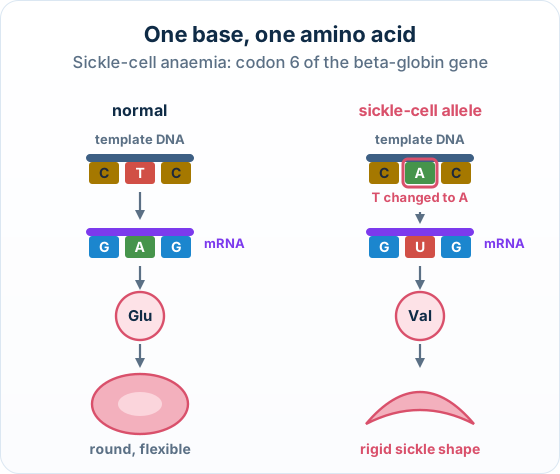
The points to remember
- Sickle-cell anaemia: one base substitution in the beta-globin gene.
- The mRNA codon changes from GAG to GUG.
- Glutamic acid is replaced by valine.
- Haemoglobin S sticks together when oxygen is low, so red blood cells become sickle-shaped.
Remember it as: GAG to GUG: Glu to Val, round to sickle.
Real example: where it is common. Sickle-cell anaemia is most common in families from parts of sub-Saharan Africa, the Middle East and India, where malaria is or was widespread.
How this is tested: The signature Paper 2 task here is a 4-mark Outline of how a single DNA base substitution leads to sickle-cell anaemia at the molecular level.
The marks: name the cascade in order
Base substitution
One DNA base is swapped.
Changed codon
One mRNA codon changes.
One amino acid changed
Glutamic acid -> valine.
Abnormal haemoglobin
The protein is altered.
Sickled cells
Red blood cells sickle.
What else the question may ask: The same long question often continues into a Punnett grid for two carrier parents (covered in monohybrid crosses) and an Explain of why the sickle allele persists in malaria regions (covered in natural selection) — but the molecular cascade is the part tested here.
Exam-style question — outline the molecular cascade
Outline how a single DNA base substitution leads to sickle-cell anaemia at the molecular level. [4]
How to score all four marks
- Start at the gene. One base is substituted in the DNA of the haemoglobin gene, creating a new allele.
- Move to the mRNA / amino acid. This changes one mRNA codon, so one amino acid in the haemoglobin chain is changed — glutamic acid is replaced by valine.
- Reach the protein. The result is abnormal haemoglobin (HbS) that sticks together into fibres when oxygen is low.
- Reach the cell. The red blood cells are pulled into a rigid sickle shape, which is the cause of sickle-cell anaemia.
Final answer
A base substitution in the haemoglobin gene changes one codon, so one amino acid changes (glutamic acid -> valine); this makes abnormal haemoglobin (HbS) that sticks together when oxygen is low, pulling red blood cells into a rigid sickle shape.
Why this scores full marks: It is a linked chain, each step causing the next — gene, then codon/amino acid, then protein, then cell.
An 'outline' worth 4 marks needs the steps in order, not four ways of saying 'one base changed'. The named swap (glutamic acid -> valine) is the detail that separates a top answer.
| Feature | Normal (HbA) | Sickle-cell (HbS) |
|---|---|---|
| The base in the gene | Original base present | One base substituted |
| The 6th amino acid in the chain | Glutamic acid | Valine |
| Behaviour when oxygen is low | Stays dissolved and flexible | Sticks together into fibres |
| Red blood cell shape | Round, flexible disc | Rigid sickle (crescent) |
| Effect on the body | Carries oxygen normally | Blocks capillaries; carries less oxygen |