Biologists use two kinds of microscope. Each one has a clear strength and a clear trade-off.
Light microscope
- Shines a beam of light through a thin sample
- Cheap and easy to use
- Can show living, moving cells
- Resolution is limited by the wavelength of light, so the smallest internal structures stay blurry
Electron microscope
- Uses a beam of electrons instead of light
- Reveals far smaller detail, such as ribosomes and the internal structure of organelles
- The sample must be killed and specially prepared
- You can never watch a living cell with it
- Light microscope
- A microscope that uses a beam of light; lower resolution, but can view living cells.
- Electron microscope
- A microscope that uses a beam of electrons; much higher resolution, but samples must be killed and treated first.
- Ultrastructure
- The fine internal detail of a cell (such as membranes and ribosomes) that only the electron microscope can reveal.
Both microscopes send a beam through the sample to form a magnified image — but the light microscope uses light (lower resolution, living cells OK) while the electron microscope uses electrons (much higher resolution, sample killed).
Interactive diagram
Explore the labelled diagram, charts and maps for this topic in full study mode.
| Feature | Light microscope | Electron microscope |
|---|---|---|
| What it uses | a beam of light | a beam of electrons |
| Resolution (detail seen) | lower — limited by the wavelength of light | much higher — can show tiny detail |
| Smallest things visible | whole cells, nuclei, chloroplasts | ribosomes, membranes, internal organelle structure |
| Living samples? | can view living, moving cells | no — samples are killed and treated first |
| Colour? | natural / stained colours | black-and-white (colour is added later) |
Why the electron microscope was such a breakthrough: Before electron microscopes, biologists could only see whole cells and the largest structures.
Because the electron microscope has much higher resolution, it revealed organelles and detailed internal structure for the first time — transforming our understanding of what a cell is made of.
Newer methods go even further: Cryogenic electron microscopy (cryo-EM) flash-freezes a sample so fast that it captures a sharp snapshot of delicate structures — even individual proteins — in close to their natural shape, without the damage that older preparation caused.
Key terms to keep straight: Light microscope → light, lower detail, living cells OK.
Electron microscope → electrons, much finer detail, samples killed.
Cryo-EM → a type of electron microscopy that freezes the sample for a snapshot of proteins and delicate structures.
Free preview
This is the free notes preview
You're reading the free notes. Aimnova Pro unlocks the full study experience — and you can try it with your first topic free to keep:
- FlashcardsLock in vocabulary and key terms with spaced repetition.
- Practice questionsAnswer exam-style questions and get instant AI marking.
- Mock exams & past-paper vaultSit full mocks and see exactly how examiners award marks.
- Personalised study planA daily plan built around your exam date and weak areas.
The big idea: Newer methods show what older ones could not: inside membranes, the shapes of single proteins, and where one kind of molecule is in a cell.
The points to remember
- Electron microscopy: a beam of electrons gives far higher resolution, showing organelles and ultrastructure.
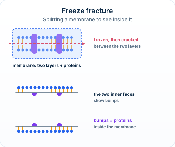
- Freeze fracture: a frozen sample is cracked along the middle of its membranes, showing their internal structure.
- Cryogenic electron microscopy (cryo-EM): proteins are flash-frozen in solution, so their shape can be imaged.
- Fluorescent stains bind to particular parts of a cell and glow in light of one colour.
- Immunofluorescence: a fluorescent stain is attached to an antibody, which binds to one antigen, showing where it is.
- Fluorescence works in a light microscope, so living cells can be watched.
Remember it as: Crack it, freeze it, light it up.
Real example: the coronavirus spike: In February 2020, weeks after the virus was identified, scientists used cryo-EM to show the shape of the spike protein of SARS-CoV-2. The structure helped scientists design vaccines.
Keep the antibody and the antigen the right way round:
The stain goes on the antibody: In immunofluorescence the fluorescent stain is attached to the antibody. The antibody then binds to the antigen, the molecule being looked for.
Get feedback like a real examiner
Submit your answers and get instant feedback — what you did well, what's missing, and exactly what to write to score full marks.
How it is tested: Paper 1A asks three things: what freeze fracture shows, which technique fits a description (often cryo-EM), and a feature of immunofluorescence. Match the method to what it reveals.
| Method | What it shows | Key word |
|---|---|---|
| Electron microscopy | Organelles and ultrastructure | higher resolution |
| Freeze fracture | The internal structure of membranes | split between the layers |
| Cryo-EM | The shape of single proteins | flash-frozen in solution |
| Immunofluorescence | Where one molecule is in a cell | stain attached to an antibody |
Outline the advantages of freeze fracture, cryogenic electron microscopy and immunofluorescence.
Model answer plan
See the answer broken into clear points, with guidance on how each point earns marks.